

Cranial and Spinal Nerves: Definition, Function, Diagram, Function, Structure
Cranial and spinal nerves form the communication network between the CNS and the rest of the body. Cranial nerves mainly serve the head and neck, while spinal nerves innervate the trunk and limbs. This guide explains their anatomy, pathways, functions, differences, NEET MCQs, clinical notes, and diagrams.
This Story also Contains
- Overview of The Nervous System
- What Are Cranial Nerves?
- What Are Spinal Nerves?
- Cranial vs Spinal Nerves — Key Differences
- Clinical & Diagnostic Importance
- Cranial & Spinal Nerves NEET MCQs (With Answers & Explanations)
- Recommended Video on "Cranial and Spinal Nerves"
Overview of The Nervous System
The nervous system forms a complex network that controls and integrates the activities of the body.
Central Nervous System (CNS)
The brain is the management centre of thoughts, emotions, and body functions.
Spinal cord is a channel for signals between the brain and the rest of the body.
Peripheral Nervous System (PNS)
The system of nerves outside the CNS is responsible for linking the CNS to limbs and organs.
Communicates between the brain/spinal cord and peripheral body parts.
What Are Cranial Nerves?
Cranial nerves are major components of the peripheral nervous system involved in sense and function activities within the head and neck.
Origin
These nerves come directly out of the brain.
There are 12 pairs of cranial nerves.
These nerves take care of the sensory and motor activities related to the head and the neck.
Classification
Sensory: Concerned with senses like smell, vision, and hearing.
Motor: Responsible for controlling muscle movements.
Mixed: They both act as sensory and motor nerves.
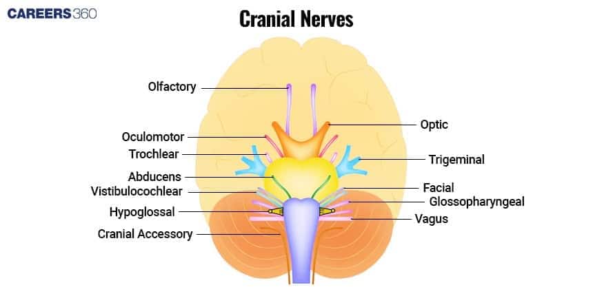
List of Cranial Nerves with Functions & Pathways
Every cranial nerve performs specific functions and takes specific pathways from the brain to the places they need to go to.
Cranial Nerve | Function | Pathway |
|---|---|---|
Olfactory Nerve | Sense of smell | Nasal cavity to olfactory bulb |
Optic Nerve | Vision | Retina to brain |
Oculomotor Nerve | Eye movement, pupil constriction | Midbrain to Eye Muscles |
Trochlear Nerve | Eye movement (superior oblique muscle) | Midbrain to eye muscles |
Trigeminal Nerve | Facial sensation, chewing muscles | Pons to face |
Abducens Nerve | Eye movement (lateral rectus muscle) | Pons to eye muscles |
Facial Nerve | Facial expression, taste (anterior 2/3 of the tongue) | Pons to face |
Vestibulocochlear Nerve | Hearing and balance | Inner ear to brain |
Glossopharyngeal Nerve | Taste (posterior 1/3 of the tongue), swallowing | Medulla to throat |
Vagus Nerve | Automatic functions of the heart, lungs and digestive tract | Medulla to organs in thorax and abdomen |
Accessory Nerve | Shoulder and neck muscles | Medulla and spinal cord to muscles |
Hypoglossal Nerve | Tongue Movement | Medulla to tongue muscles |
Common Disorders
Conditions like Bell's palsy, trigeminal neuralgia, and anosmia reflect the clinical relevance of cranial nerves.
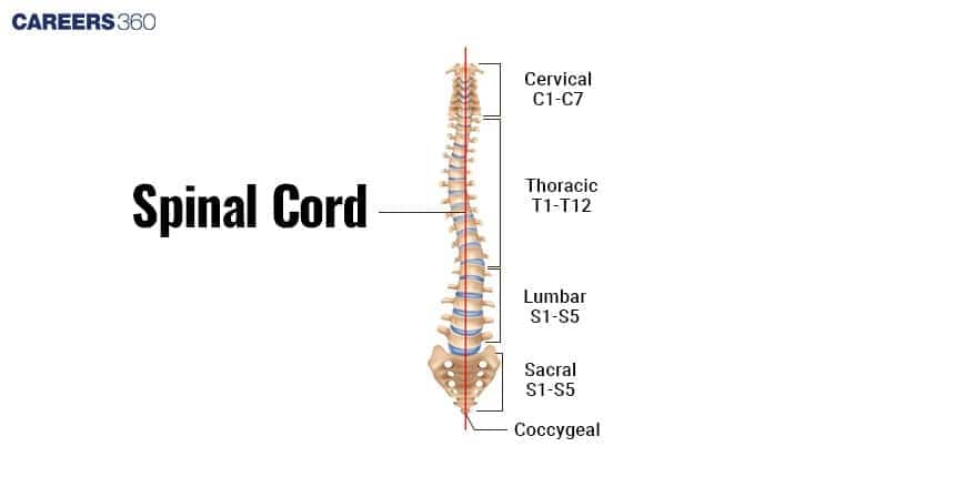
What Are Spinal Nerves?
Spinal nerves are essential for the transmission of signals between the spinal cord and the body.
Origin
These nerves have their origin in the spinal cord.
The number of spinal nerves is thirty-one pairs.
Anatomy Of Spinal Nerves
Root and rootlets: Nerve roots which further subdivide into rootlets.
Dorsal and ventral roots: Dorsal roots transmit sensory signals, while the ventral roots transmit motor signals.
Functions And Pathways
Branches of the spinal nerves innervate the body and carry out sensory and motor functions.
Divisions of Spinal Nerves
| Division | Number of Pairs |
|---|---|
Cervical | 8 pairs |
Thoracic | 12 pairs |
Lumbar | 5 pairs |
Sacral | 5 pairs |
Coccygeal | 1 pair |
Common Disorders
Sciatica, herniated disks, and spinal stenosis are conditions that affect the spinal nerves. Comparisons:
Cranial vs Spinal Nerves — Key Differences
Key differences between cranial and spinal nerves is discussed in the table below:
| Cranial Nerves | Spinal Nerves | |
|---|---|---|
Origin | Emerge from the brain | Emerge from the spinal cord |
Types of fibres | May be sensory, motor, or mixed | All are mixed (sensory + motor) |
Target Regions | Supply head and neck (except vagus) | Supply trunk and limbs |
Number | Twelve Pairs | Thirty-one pairs |
Pathway Complexity | More specialized pathways | More uniform segmental distribution |
Clinical & Diagnostic Importance
The knowledge of cranial and spinal nerves is crucial in medicine concerning the diagnosis and treatment of neurological disorders within these nerves.
Medicinal Importance:
Nerve functions and pathways play a significant role in the diagnosis and treatment of diseases.
Diagnostic Measures:
MRI: It details the structure of nerves.
CT scans: They help identify any structural abnormalities.
Electromyography: It is a test to measure the electrical activities in the muscles, so the test helps in diagnosing nerve damage.
Treatment:
Medications, physical therapy, and surgical interventions are the treatment options for nerve disorders.
Cranial & Spinal Nerves NEET MCQs (With Answers & Explanations)
Important questions asked in NEET from this topic are:
Functions of cranial and spinal nerves
Cranial vs Spinal Nerves
Practice Questions for NEET
Q1. Identify the suitable option,
Assertion: Trigeminal neuralgia or douloureux is a clinical condition which causes severe pain in the face.
Reason: Cranial nerve V or trigeminal nerve when compressed at the root entry by the superior cerebellar artery causes Trigeminal neuralgia
The reason is correct for the assertion.
The reason is not correct.
Reason and assertion both are incorrect.
The reason is correct but the assertion is incorrect.
Correct answer: 1) The reason is correct for the assertion.
Explanation:
A clinical disorder known as trigeminal neuralgia is characterized by acute, electrical, shock-like, paroxysmal lancinating pain in the region served by one or more trigeminal nerve branches. The superior cerebellar artery compresses the trigeminal nerve at the root entrance zone in the majority of cases. The chronic pain condition known as trigeminal neuralgia (TN), commonly referred to as tic douloureux, causes abrupt, excruciating face agony. The fifth cranial nerve, the trigeminal nerve, which supplies feeling and nerve communication to several areas of the head and face, is impacted.
Hence, the correct option is (1)The reason is correct for the assertion.
Q2. Cranial nerve which shows maximum branching is
Trigeminal
Vagus
Optic
Facial
Correct answer: 1) Trigeminal
Explanation:
This is the biggest cranial nerve, and the most extensive branching of which is the trigeminal nerve or cranial nerve V. This has three major branches.
Ophthalmic Nerve (V1): It is a sensory innervation of the forehead, upper eyelid, and parts of the nasal cavity.
Maxillary Nerve (V2): The nerve provides sensory fibers for the cheeks, upper lip, and upper teeth.
Mandibular Nerve (V3): The largest branch, which carries sensory as well as motor fibers supplying the lower jaw and musculature of mastication.
Its intricate anatomy allows the trigeminal nerve to undertake vast sensory and motor functions all over the face, therefore being important both in sensation of the face and mastication.
Hence, the correct answer is option 1) Trigeminal.
Q3. Injury to the vagus nerve in humans is not likely to affect : (Concept - Cranial Nerves)
Tongue movements
Gastrointestinal movements
Pancreatic secretion
Cardiac movement
Correct answer: 1) Tongue movements
Explanation:
The vagus nerve (cranial nerve X) regulates the heart rate, digestion, and speech patterns, among other things. It also contributes to swallowing and voice production by innervating the pharyngeal and laryngeal muscles. It does not, however, regulate tongue motions. Both the intrinsic and extrinsic muscles of the tongue are innervated by the hypoglossal nerve (cranial nerve XII), which is principally responsible for controlling tongue motions. The vagus nerve would therefore not be directly impacted by damage to the tongue.
Hence, the correct answer is option 1)Tongue movements.
Also Read:
Recommended Video on "Cranial and Spinal Nerves"
Frequently Asked Questions (FAQs)
Diagnostics include MRI, CT scans, and electromyography to assess structural and functional abnormalities.
Twelve cranial nerves: Olfactory, Optic, Oculomotor, Trochlear, Trigeminal, Abducens, Facial, Vestibulocochlear, Glossopharyngeal, Vagus, Accessory, and Hypoglossal.
Spinal nerves carry sensory and motor signals between the spinal cord and the rest of the body, controlling movement and sensation in the trunk and limbs.
Disorders like Bell's palsy, trigeminal neuralgia, sciatica, and herniated discs will affect these nerves, resulting in pain, loss of function, or sensory deficits.
Cranial nerves deal with the sensory and motor functions of the head and neck, including smell, vision, taste, hearing, and movements of the face.
