Choroid plexus: Definition, Function, Structures, Diagram, Facts, FAQs
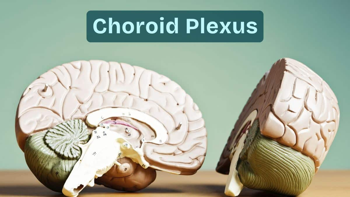
The choroid plexus is a specialised brain structure that produces cerebrospinal fluid (CSF) and maintains the blood–CSF barrier. Located in all four ventricles, it plays a vital role in regulating the brain’s internal environment. This guide covers its anatomy, histology, functions, diagrams, NEET notes, FAQs, and MCQs.
This Story also Contains
- What Is the Choroid Plexus?
- Anatomy of The Choroid Plexus
- Histology of the Choroid Plexus
- Functions of The Choroid Plexus
- Clinical Importance
- Choroid Plexus NEET MCQs (With Answers & Explanations)
- Recommended video for "Choroid Plexus"

What Is the Choroid Plexus?
The choroid plexus is a collection of cells located within the ventricles of the brain, especially inside the lateral, third, and fourth ventricles. It comprises ependymal cells, blood vessels, and connective tissue and forms the CSF. The choroid plexus filters blood for the production of CSF, which later goes into the brain and spinal cord, cushioning, nourishing, and clearing waste products. It forms a part of the maintenance of the internal environment of the brain and helps in the sustenance of neural health for overall correct functionality of the central nervous system.
Anatomy of The Choroid Plexus
The choroid plexus is a structure inside the ventricles of the brain that accounts for the largest proportion of cerebrospinal fluid production. It can be found in all parts of the ventricles inside the brain, namely the lateral, third, and fourth ventricles.
Location in the Brain Ventricles
The choroid plexus lies in all four ventricles of the brain, namely, two lateral ventricles, the third ventricle, and the fourth ventricle.
The location extends from the lateral ventricles through the interventricular foramina into the third ventricle, then goes on through the cerebral aqueduct into the fourth ventricle.
Structural Components
The choroid plexus is composed of an epithelium layer, connective tissue, and a rich network of blood vessels.
The epithelium is lined by cuboidal choroidal epithelial cells that secrete CSF.
The choroidal epithelial cells are underlaid by thin stroma connective tissue under the epithelium to which blood vessels that supply the plexus are anchored.
These blood vessels are fenestrated and allow the filtration of blood plasma into the ventricles to form CSF.

Histology of the Choroid Plexus
The choroid plexus is very defined and complicated when viewed microscopically.
Ependymal / Choroidal Epithelial Cells
These epithelial cells, otherwise named ependymal cells, appear cuboidal to columnar and cover the connective tissue core underlying them continuously.
The major cells of the choroid plexus include epithelial cells, also called choroidal epithelial cells or ependymal cells.
These cells have microvilli and cilia on the apical surface that will aid in the movement and secretion of CSF.
Stroma & Vascular Network
Under the stromal connective tissue, there are several kinds of support cells and blood vessels.
They help in the exchange of materials for CSF production.
Functions of The Choroid Plexus
The choroid plexus is the part of the brain that is responsible for the significant production of CSF and the maintenance of the blood-CSF barrier.
CSF Production
CSF is produced by the choroid plexus through the process involving filtration of blood plasma.
This makes the blood vessels within the choroid plexus fenestrated, and as such, plasma passes through them into the stroma.
At this point, the choroidal epithelial cells secrete this filtrate into the ventricles as CSF, enriched with essential ions, nutrients, and other substances.
With this, it controls the intracranial pressure and gives the best environment for the proper functioning of the neurons.
Regulation of CSF Volume & Composition
It regulates the volume and composition of the CSF.
It maintains the volume of the CSF with the proper balance of the electrolytes, glucose, and other essential molecules.
Formation of the Blood–CSF Barrier
The blood-CSF barrier derives from tight junctions between the choroidal epithelial cells.
These junctions hamper free diffusion into the CSF from the blood for large molecules and pathogens but allow, in a selective manner, the majority of essential nutrients and ions.
Neuroprotection
Blood-CSF prevents most of the noxious chemicals and pathogenic agents from the blood into the CSF and eventually into the CNS.
It becomes rather important to maintain such an optimal environment to protect the safe and stable operation of the brain.
Clinical Importance
The clinical importance of choroid plexus includes:
Hydrocephalus (overproduction/blockage): Hydrocephalus occurs when CSF is overproduced or its flow is blocked, leading to increased pressure inside the skull and potential brain damage.
CSF leakage: Leakage of cerebrospinal fluid reduces brain cushioning and can cause severe headaches, infections, and low-pressure complications.
Infections and barrier failure: When pathogens cross the blood–brain or CSF barriers, serious infections like meningitis can develop due to loss of protective filtration.
Choroid Plexus NEET MCQs (With Answers & Explanations)
Important questions asked in NEET from this topic are:
Anatomy of choroid plexus
Functions of choroid plexus
Practice Questions for NEET
Q1. Cerebrospinal fluid is secreted by
Choroid plexus
Foramina of Monro
Foramen of Magendie
Cauda equina
Correct answer: 1) Choroid plexus
Explanation:
Cerebrospinal fluid (CSF) is a colorless, transparent liquid produced by choroid plexuses, which are created from vascular tissue situated in the brain's ventrals. A CSF is produced by the choroid plexus made up of a capillary bed and ependymal cells that extract blood plasma and filter it. This fluid is moved among the subarachnoid space, spinal cord central canal, and ventricles.
Hence, the correct answer is option 1) Choroid plexus.
Q2. Cerebrospinal fluid fills the space between
The arachnoid and pia mater of the central nervous system (CNS)
The ventricles of the brain
The central canal of the spinal cord
All of the above
Correct answer: 4) All of the above
Explanation:
The cerebrospinal fluid (CSF) is a clear watery nutritional fluid containing sodium chloride, potassium, glucose, and various proteins. It fills the space between the arachnoid and pia mater of the central nervous system (CNS), the ventricles of the brain, and the central canal of the spinal cord. It is secreted by the choroid plexuses. In addition to providing a nutritional supply to the brain, the CSF also removes by-products of metabolism and also serves to provide a fluid cushion to protect the brain from injury.
Hence, the correct answer is option 4) All of the above.
Q3. Assertion: CSF Fluid movement is pulsatile, matching the pressure waves generated in blood vessels by the beating of the heart.
Reason: CSF moves in a single outward direction from the ventricles, but multi directionally in the subarachnoid space.
If both Assertion & Reason are true and the reason is the correct explanation of the assertion, then mark A
If both Assertion & Reason are true but the reason is not the correct explanation of the assertion, then mark B
If Assertion is true statement but Reason is false, then mark C
If both Assertion and Reason are false statements, then mark D
Correct answer: 1) If both Assertion & Reason are true and the reason is the correct explanation of the assertion, then mark A
Explanation:
Both Assertion & Reason are true and the reason is the correct explanation of the assertion. CSF moves in a single outward direction from the ventricles, but multi directionally in the subarachnoid space. Fluid movement is pulsatile, matching the pressure waves generated in blood vessels by the beating of the heart. But believes that there is no unidirectional CSF circulation, but cardiac cycle-dependent bi-directional systolic-diastolic to-and-from cranio-spinal CSF movements.
Hence, the correct answer is option 1) If both Assertion & Reason are true and the reason is the correct explanation of the assertion, then mark A
Also Read:
Recommended video for "Choroid Plexus"
Frequently Asked Questions (FAQs)
The most vital role played by the choroid plexus is that it fabricates and controls cerebrospinal fluid.
It is found in the ventricles of the brain. The choroid plexus is situated within the lateral ventricles, third ventricle, and fourth ventricle.
The disorders related to it are hydrocephalus, the Choroid plexus Papillomas/ Carcinomas. Other conditions include infections and inflammations of the choroid plexus, thus affecting CSF formation and composition.
Imaging techniques such as magnetic resonance imaging and computed tomography show the anatomical structure of the choroid plexus and define any abnormality.
Yes, a few potential uses of choroid plexus cells are in the pipeline in regenerative medicine that is currently under research for neurological disorders and brain injuries.
