

Difference Between Arteries and Veins
Arteries and veins are the two core blood vessels maintaining circulation by transporting oxygenated and deoxygenated blood between the heart and body. Their differences in structure, pressure levels, lumen size, and valves form the basis of NEET-level vascular physiology. A clear comparison helps master circulation disorders, portal system concepts, and exam-grade MCQs.
This Story also Contains
- What are Arteries and Veins?
- Structure of Arteries And Veins
- Types of Arteries
- Types of Veins
- Characteristics of Arteries and Veins
- Key Differences between Arteries and Veins
- Arteries vs Veins NEET MCQs (With Answers & Explanations)
- Recommended Video on Difference Between Arteries and Veins
What are Arteries and Veins?
Blood vessels are part of the circulatory system which includes arteries and veins. These blood vessels transport blood all over the body and capillaries. Arteries are blood vessels that transport well-oxygenated blood from the heart to different parts of the body whereas veins are the vessels that transport poorly oxygenated blood back to the heart. Both of these types of vessels are essential for adequate blood flow, the delivery of oxygen and nutrients to the tissues and the removal of waste products from these tissues. An overview of arteries and veins is therefore important in understanding the concept of circulatory systems and the significance of vascular health.
Structure of Arteries And Veins
The structure of arteries and veins is discussed below:
Structure of Arteries
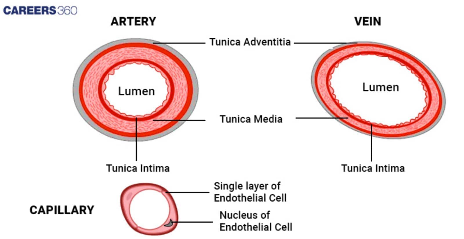
Thick walls: Arteries have thick walls made up of three tunics. Tunica intima, which is the inner layer of flat endothelial cells, tunica media which is a muscular and elastic layer found in large arteries, and tunica externa the outermost connective layer. Large muscular walls guard and control high blood pressure resulting from the blood pumped from the heart.
Elastic fibres: These are required to give proper flexibility to arteries for each contraction and relaxation during the heartbeat to manage proper blood flow.
Narrow lumen: The arteries hence have a relatively small inner diameter known as the lumen hence the ability to withstand high pressure to enable blood to flow nicely.
Smooth muscle cells: These are located in the tunica media control the contraction and dilation of arteries to control blood pressure and the rates of flow of blood in the body.
Structure of Veins
Thin walls: Compared to the arteries, veins are vessels of smaller diameter, they do not have much musculature and elastic tissue because they pump the blood under lower pressure.
Less elastic tissue: The elasticity is less in the veins this is because veins return blood at low pressure to the heart.
Wide lumen: Veins are blood vessels with slightly wider lumen to contain a significantly larger amount of blood that will be pumped back to the heart.
Valves: Veins that are found in the legs part have valves which prevent the backflow of blood and the blood will only flow in one direction to the heart.
Types of Arteries
The types of arteries are:
Elastic Arteries
These are the largest arteries nearest to the heart and they have thick walls with greater proportionality of elastic fibres. This elasticity enables them to receive blood when there is an increase in pressure as a result of pumping from the heart and ensures normal flow of blood.
Examples: Pulmonary artery and the aorta.
Muscular Arteries
These arteries contain a proportionately larger quantity of smooth muscles rather than elastic fibres, through which they can regulate the blood flow by changing the diameter of the blood vessels. They transport blood to other organs of the body.
Examples: The radial artery and the femoral artery.
Arterioles
They are tiny blood vessels that regulate the quantity of blood to be delivered to an area of the body. It includes a rather feeble muscular layer compared to its measures and they have chances to control blood flow discreetly and alter the pressure.
Function: They act as pressure maintaining and directing vessels whereby they maintain blood pressure and then direct the circulations into capillary beds.
Types of Veins
The types of veins are:
Superficial Veins
These veins are near the dermis and therefore they are easily seen on the skin. They allow blood and fluid to be drawn out of the skin and the first layers of the tissue.
Examples: The greater saphenous vein and the small saphenous vein.
Deep Veins
Located more towards the centre and frequently in association with arteries, deep veins transport the blood from internal organs and muscles and back to the heart. They are more crucial for blood flow and pressure maintenance.
Examples: The femoral vein provided the major contribution to the popliteal vein while the brachial vein contributed to the antebrachial vein.
Venules
These are the smallest blood veins and take blood deposits from the tiny capillaries and these accumulate in other bigger veins. It is thin-walled and is engaged in the process of collecting blood from the capillary beds.
Function: Capillaries are small blood vessels which allow blood to flow from arterioles to venues and thus play a very important role in circulation by channeling blood back to the veins.
Characteristics of Arteries and Veins
The characteristics of arteries and veins is discussed below:
Characteristics of Arteries
1. Situated deep into a muscle
2. Have extremely thick walls
3. Transfer blood between the organs and the heart.
4. Deliver oxygen-rich blood (except for the pulmonary artery)
5. Internally has a thick layer of muscular tissue
6. Lack valves (except for the pulmonary artery)
Characteristics of Veins
1. Are situated closer to your body's surface.
2. Possess thin walls
3. Direct blood flow to your heart
4. Carry anaemic blood
5. Contain a thin coating of muscular tissue
6. Have valves to maintain blood flow
Key Differences between Arteries and Veins
It is one of the important difference and comparison articles in biology. The differences are listed below:
Aspect | Arteries | Veins |
Wall Thickness | Thick, muscular walls | Thin walls |
Lumen Diameter | Narrow lumen | Wider lumen |
Presence of Valves | No valves (except in the aorta and pulmonary arteries) | Valves present to prevent backflow |
Elasticity and Muscularity | High elasticity and muscularity | Less elasticity and muscularity |
Blood Oxygenation Levels | Typically oxygen-rich (except pulmonary arteries) | Typically oxygen-poor (except pulmonary veins) |
Pressure Levels | High pressure due to pumping from the heart | Low pressure as blood returns to the heart |
Blood Flow Direction | Away from the heart to various tissues | Toward the heart from the tissues |
Arteries vs Veins NEET MCQs (With Answers & Explanations)
Types of questions asked from this topic are:
Types of arteries and veins
Difference between arteries and veins
Practice Questions for NEET
Q1. Which of the following accurately describes a portal system in the human body?
A vein originates from an organ and terminates in the heart.
A vein enters an organ other than the heart and branches into capillaries.
An artery terminates in an organ and restarts through the fusion of its capillaries.
Blood from the intestine is routed through the kidneys before reaching the inferior vena cava (IVC).
Correct answer: 2) A vein enters an organ other than the heart and branches into capillaries
Explanation:
In a portal system, a special type of vein carries blood from one organ to another without first going to the heart. Instead, it directly enters a specific organ, like the liver or hypothalamus. Inside that organ, the vein branches out into many tiny blood vessels called capillaries, forming a network. This arrangement allows the organ to perform specific tasks like filtering substances, metabolizing nutrients or regulating hormones. After these functions are carried out, the blood then continues its circulation to other parts of the body. So, in a portal system, the blood takes a detour through a secondary organ before returning to the heart.
Hence, the correct answer is option 2) A vein enters an organ other than the heart and branches into capillaries.
Q2. Which of the following is true?
All arteries carry oxygenated blood
All veins carry deoxygenated blood
All arteries carry oxygenated blood except one
All veins carry oxygenated blood except one
Correct answer: 3) All arteries carry oxygenated blood except one
Explanation:
Arteries
These are elastic vessels that transport blood away from the heart
The largest artery of the body is the aorta
The aorta originates from the heart and branches out into smaller arteries.
The smallest branches are called arterioles, which further branch off into capillaries
So, All arteries carry oxygenated blood except pulmonary arteries.
Hence, the correct answer is option 3) All arteries carry oxygenated blood except one.
Q3. In veins thickest tunica is,
Tunica media
Tunica Externa
Tunica interna
Both a and b
Correct answer: 2) Tunica Externa
Explanation:
In veins thickest tunica is, tunica externa as it provides structural integrity and attachment to surrounding tissues. Even when blood pressure is low, the tunica externa is essential for maintaining the shape of the vein and preventing it from collapsing.
Hence, the correct answer is option 2)Tunica Externa.
Also Read:
Recommended Video on Difference Between Arteries and Veins
Frequently Asked Questions (FAQs)
Arteries: Plaque deposition, aneurysms and hypertension
Veins: Varicose veins, deep vein thrombosis, and chronic venous insufficiency
Arteries have thick walls to withstand the pressure resulting from the blood that is ejected out of the heart. Due to the presence of thick muscular and elastic layers blood pressure is maintained and blood is easily pumped through the arteries.
Antegrade blood flow is always in the arteries; its oxygenation is typically high except in the pulmonary arteries. The dark-coloured blood contains lesser oxygless returns from the tissues back to the heart mainly through the veins.
