

Heart Pump Of The Circulatory System
The human heart is a muscular organ that maintains blood flow and supports oxygen and nutrient transport throughout the body. It includes four chambers, valves, a conduction system, and a rhythmic cardiac cycle ensuring continuous circulation. This guide explains heart structure, cardiac cycle, valve function, conduction pathway, NEET notes, and exam-focused MCQs.
This Story also Contains
- What Is Human Heart?
- Basic Anatomy of the Human Heart
- Blood Circulation Through the Heart
- Cardiac Cycle (Systole & Diastole)
- Heart Valves and Their Function
- Electrical Conduction System of the Heart
- Regulation of Heartbeat (ANS + Hormones)
- Blood Vessels and Circulation Types
- Cardiovascular Health and Disorders
- Heart NEET MCQs (With Answers & Explanations)
- Recommended Video on the Functioning of the Human Heart
What Is Human Heart?
The human heart is an organ beyond compare, serving as the central pump for the circulatory system. This heartbeat, beating over 30 million times in a year, supplies the tissues and organs of the human body with blood, oxygen, and nutrients.
Basic Anatomy of the Human Heart
The anatomy of the heart is discussed below:
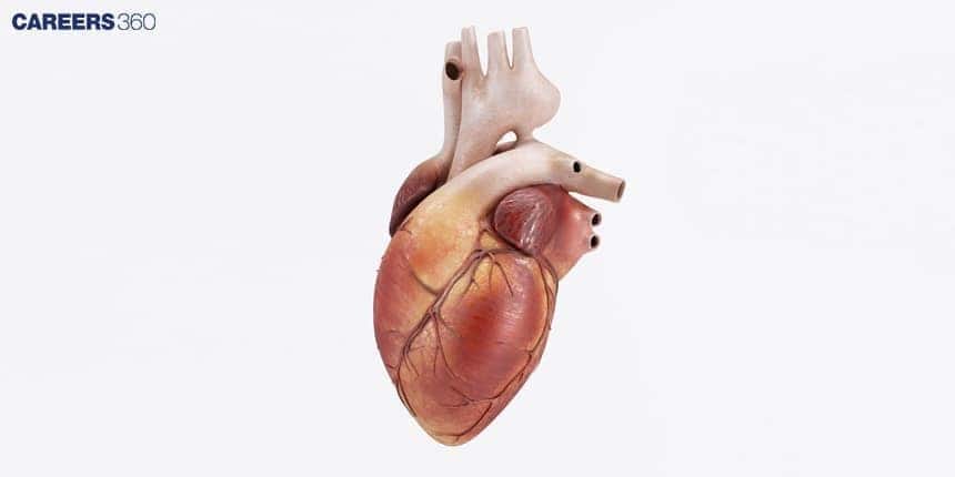
Location & Pericardium
The heart is a two-sided, four-chambered muscular pump positioned in the thoracic cavity, flanked by the lungs. Enclosed is a two-layered covering called the pericardium. The four chambers of the heart, from superior to inferior, include the right and left atria and the right and left ventricles.
Four Chambers
The chambers are separated by walls of muscle. The two upper chambers are called the atria, or atrium singular, and have thin walls and small, protruding appendages called auricles. The interatrial septum isolates the right atrium from the left atrium.
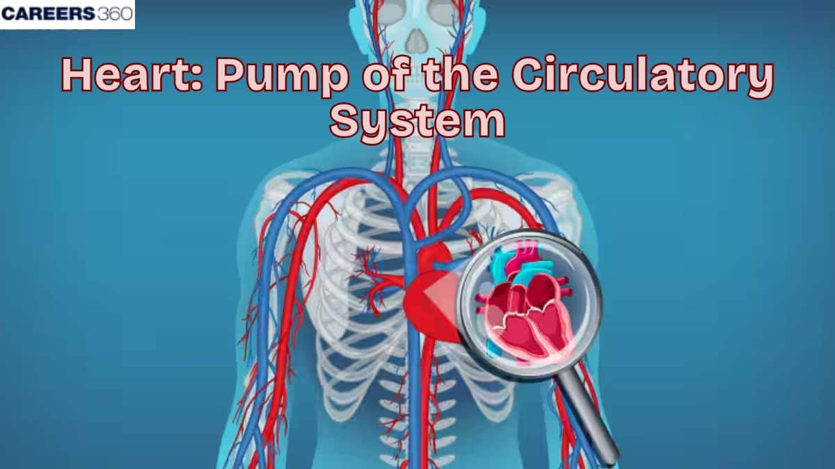
Blood Circulation Through the Heart
Blood is pumped in a circulatory pathway. Blood that is deoxygenated from the body enters the right atrium via the superior and inferior vena cava. Blood then moves to the right ventricle, which is further pumped to the lungs to be oxygenated through the pulmonary arteries.
The oxygenated blood returns to the left atrium through the pulmonary veins and then moves on to the left ventricle. The left ventricle then pumps this oxygenated blood throughout the body via the aorta. With this circulation, the blood effectively transports enough oxygen and nutrients to all body tissues while, at the same time, removing carbon dioxide and wastes.
Cardiac Cycle (Systole & Diastole)
The cardiac cycle essentially comprises alternating two main phases: diastole and systole. Diastole is the period when the heart chambers are relaxed and are filled with blood. On the other hand, systole is the period when heart muscles contract and force the blood out of the heart chambers.
However, the moment the contraction is over, blood begins to flow into the heart chambers and the cycle continues. The graphs of pressure changes in the heart over the duration during which the cardiac cycle occurs depict these phases and how they coordinate themselves.
Heart Valves and Their Function
Heart valves are structures, which facilitate the flow of blood in one direction. The right and left atria are prevented from backflow when the closing of the aortic and pulmonary valves is done during the phase of diastole. The opening and closing of the valves are maintained by the changes in pressure developed in the chambers of the heart.
Atrioventricular Valves
Because they are located between an atrium and a ventricle, the tricuspid and bicuspid valves are termed atrioventricular (AV) valves. When placed between the atrium and the ventricle, the tricuspid valve avoids the regurgitation into the right atrium.
Blood passes from the left atrium into the left ventricle through the bicuspid (mitral) valve, as its name implies it has two cusps. Blood passes from the right atrium into the right ventricle through a valve that is called the tricuspid valve because it consists of three cusps. It is also called the right atrioventricular valve.
Semilunar Valves
The aortic and pulmonary valves are known as the semilunar (SL) valves because they are made up of three crescent moon–shaped cusps. Each cusp attaches to the arterial wall by its convex outer margin.
Electrical Conduction System of the Heart
The precociously developed electrical conduction system of the heart paces the beat of the heart. The sinoatrial (SA) node, located in the right atrium, is the source of the heart's normal action impulses. The action potential is transmitted first to the atrioventricular (AV) node, then the bundle of His, and then out to the Purkinje fibres, enabling the spread of the impulse to the muscles in the heart to cause them to contract.
ECG (Electrocardiogram)
As action potentials propagate through the heart, they generate electrical currents that can be detected at the surface of the body. An electrocardiogram (ECG) is a recording of these electrical signals. The ECG is a composite record of action potentials produced by all of the heart muscle fibers during each heartbeat. The instrument used to record the changes is an electrocardiograph.
Regulation of Heartbeat (ANS + Hormones)
The autonomic nervous system and hormones that regulate the heartbeats. At any prospect of stress to the body, or either during any physical activity, the system of the sympathetic nerves can speed up the rate of the heartbeat, while the parasympathetic system can slow the rate down during relaxation. The presence of other hormones in the body, like adrenaline, majorly plays a role in regulating the heart rate.
Blood Vessels and Circulation Types
The circulatory system consists of arteries, veins, and capillaries. Arteries carry oxygenated blood away from the heart, veins transport deoxygenated blood back to the heart, and capillaries enable the exchange of gases, nutrients, and wastes at the tissue level.
Cardiovascular Health and Disorders
A healthy lifestyle is one of the most important ways to maintain a healthy heart. Regular exercise, a balanced diet, avoidance of cigarettes, and stress control are the norms. Protection and timely treatment are essential for maintaining heart health.
Among the most common heart diseases are:
Coronary Artery Disease: A condition that affects the blood vessels called the coronary arteries leading into the heart muscle and which get blocked thus limiting the amount of blood that is delivered to the muscle.
Heart Attack: This happens when a section of the heart does not get an adequate amount of blood and nutrients hence becoming damaged.
Heart Failure: A state in which the heart is not able to pump blood as it should, hence accumulating fluids within the body.
Heart NEET MCQs (With Answers & Explanations)
Types of questions asked from this topic are:
Anatomy of Human heart
Electrical conduction system
Disorders of the Circulatory System
Practice Questions for NEET
Q1. The human heart is derived from
Ectoderm
Mesoderm
Endoderm
Both 1 and 3
Correct answer: 2) Mesoderm
Explanation:
Early embryonic mesoderm gives rise to the heart through a region known as the cardiogenic mesoderm. Initially, the heart is shaped as two separate tubes which merge to create a single tubular structure. This tube undergoes folding and dividing processes to develop into the atria and ventricles, along with other heart components. The mesoderm is the primary origin of the heart's tissue and structure during embryonic development.
Hence, the correct answer is option 2) Mesoderm
Q2. The right aortic arch is found in
Mammals
Mammals and reptiles
Birds
Amphibians
Correct answer: 2) Mammals and reptiles
Explanation:
The aortic arch, the arch of the aorta or the transverse aortic arch is a part of the aorta between the ascending and descending aorta. The arch travels backwards so that it ultimately runs to the left of the trachea.
The right-sided aortic arch is a rare anatomical variant in which the aortic arch is on the right side rather than on the left.
Hence, the correct answer is Option (2) Mammals and reptiles
Q3. The pericardium and the pericardial fluid help in
Protecting the heart from friction, and shocks and keeping it moist
Pumping the blood
Receiving the blood from various parts of the body
None of these
Correct answer: 1) Protecting the heart from friction, and shocks and keeping it moist
Explanation:
The pericardium is a double-layered membrane surrounding the heart, consisting of an outer fibrous layer and an inner serous layer. It protects the heart, anchors it within the chest cavity, and prevents overexpansion during blood volume changes. The pericardial fluid, found between the serous layers, acts as a lubricant to reduce friction as the heart beats. This fluid also helps distribute mechanical forces evenly across the heart. Together, the pericardium and pericardial fluid ensure the heart functions efficiently within a stable and protective environment.
Hence, the correct answer is option 1) Protecting the heart from friction, and shocks and keeping it moist.
Also Read:
Recommended Video on the Functioning of the Human Heart
Frequently Asked Questions (FAQs)
Lifestyle behaviours leading to a healthier heart include proper diet, good exercise, cessation of smoking, controlled stress, and appropriate weight maintenance.
An electrocardiogram monitors the electrical activity of the heart. It is used in the diagnosis of various heart conditions that detect irregularities in heart rhythm.
The major function of the human heart is the pumping of blood through the body, thereby providing tissues with oxygen and nutrients and carrying away carbon dioxide and other wastes.
The unidirectional flow of the blood is performed by the heart with the help of the action of its four valves: tricuspid, pulmonary, mitral and last but not least, the aortic valves open and then close with the change in excepted pressure.
The SA node, also called the sinoatrial node, is the heart's natural pacemaker. This node is responsible for generating electrical impulses that establish a regular rhythm for heartbeats.
