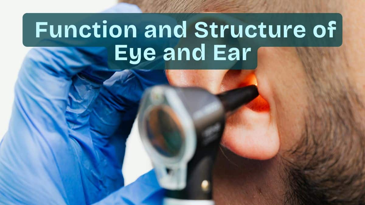
functions-and-structure-of-eye-and-ear: Structure, Function, Parts
The eye and ear are two advanced sensory organs responsible for vision and hearing—allowing humans to perceive colours, sounds, and maintain balance. Their structures include external and internal components that protect, receive, and process sensory information. This guide covers the anatomy, functions, differences, diagrams, and NEET-focused MCQs on the eye and ear together.
This Story also Contains
- Introduction — The Human Eye and Ear as Sensory Organs
- Structure of the Eye
- Structure of the Ear
- Comparison of Eye and Ear
- Eye & Ear NEET MCQs (With Answers & Explanations)
- Recommended Video on "Structure of Eye and Ear"

Introduction — The Human Eye and Ear as Sensory Organs
The human body is one such masterpiece of biological engineering where complex systems work together to keep the organism alive and allow him or her to interact with the environment. Of all such enigma-filled systems, the sensory organs are the most fascinating, especially the eye and the ear.
These organs mean colours at sunset, the soothing sound of music, or balance while walking. The structures enhance appreciation for the functioning and make a person realize just how delicate and sophisticated the sensory systems are. The anatomy of the eye and the ear details individual parts and how they interrelate to their functioning and just how much these organs are in our lives daily.
Structure of the Eye
The human eye is an extremely complex organ with various parts that assist in vision.
External Structures
These external structures not only protect the eye but also provide support in carrying out its function.
Eyelids
It protects the eye from dust and regulates the entry of light into the eye.
Conjunctiva
This is a thin membrane covering the front of the eye and lining the inside of the eyelids.
Internal Structures
These are responsible for vision.
Cornea
The transparent curved front surface of the eye helps in changing the direction of light in the eye.
Lens
A flexible, transparent structure that focuses light onto the retina.
Retina
The innermost layer comprises photoreceptors—rods and cones—that detect light and transmit the signals to the brain.
Structure of the Ear
The ear is divided into three portions: the outer ear, the middle ear, and the inner ear.
Outer Ear
That part of the ear that can be seen, collects and directs sound waves into the ear canal.
Pinna
That part of the ear is visible to our eyes.
Ear Canal
Carries the sound waves to the eardrum.
Middle Ear
The part that amplifies the sound vibrations and leads them into the inner ear.
Eardrum
Vibrates in response to the sound.
Ossicles
Small bones that can amplify sound.
Inner Ear
The part that changes sound vibrations into nerve impulses. It also maintains balance.
Cochlea
Changes sound vibrations into electrical signals.
Semicircular Canals
They maintain balance.
Comparison of Eye and Ear
The comparison between eye and ear:
| Feature | Eye | Ear |
|---|---|---|
Function | Hearing and balance | |
Major Parts | Cornea, lens, retina | Pinna, ossicles, cochlea |
Sensory Cells | Rods and cones | Hair cell |
Stimulus | Light | Sound waves and head movements |
Location | Eye socket | Temporal bone |
Eye & Ear NEET MCQs (With Answers & Explanations)
Important questions asked in NEET from this topic are:
Structure of the human eye and ear
Human ear vs eye
Practice Questions for NEET
Q1. The function of the pinna is to
helps prevent bacterial growth
collects the vibrations in the air which produce sound.
increase the efficiency of transmission of sound waves to the inner ear.
equalize the air pressure in the tympanic cavity with that on the outside.
Correct answer: 1) collects the vibrations in the air which produce sound.
Explanation:
The tympanic membrane, external auditory meatus (canal), and pinna make up the external ear. The pinna gathers the air vibrations that generate sound. It is composed of skin-covered elastic cartilage.
Hence, the correct answer is option 2) The function of the pinna is to collect the vibrations in the air which produce sound.
Q2. The kind of tissue that forms the supportive structure in our pinna (external ears) is also found in:
Nails
Ear ossicles
Tip of the nose
Vertebrae
Correct answer: 1) Tip of the nose
Explanation:
There are three types of cartilage: hyaline cartilage, fibrous cartilage, and calcified cartilage. Fibrous cartilage has well-developed fibres in its matrix and can be further classified into two types fibrous cartilage which has white strong fibres, and is located in intervertebral discs and pubic symphysis, and yellow elastic fibrous cartilage with yellow fibres that provide elasticity and is located in the ear pinna and the tip of the nose.
Hence, the correct answer is option 3) Tip of the nose.
Q3. Assertion: The outer ear (external ear) is also called the auricle or pinna, your outer ear consists of ridged cartilage and skin, and it contains glands that secrete ear wax.
Reason: Its funnel-shaped canal leads to your eardrum or tympanic membrane.
If both Assertion & Reason are true and the reason is the correct explanation of the assertion, then mark A
If both Assertion & Reason are true but the reason is not the correct explanation of the assertion, then mark B
If Assertion is true statement but Reason is false, then mark C
If both Assertion and Reason are false statements, then mark D
Correct answer: 1) If both Assertion & Reason are true and the reason is the correct explanation of the assertion, then mark A
Explanation:
Both Assertion & Reason are true and the reason is the correct explanation of the assertion. Your outer ear is the part of your ear that’s visible. It’s what most people mean when they say “ear.” Also called the auricle or pinna, your outer ear consists of ridged cartilage and skin, and it contains glands that secrete ear wax. Its funnel-shaped canal leads to your eardrum or tympanic membrane.
Hence, the correct option is 1 both Assertion & Reason are true and the reason is the correct explanation of the assertion.
Also Read:
Recommended Video on "Structure of Eye and Ear"
Frequently Asked Questions (FAQs)
The cochlea transforms the vibrations of sound into an electrical signal.
The ossicles amplify the vibrations of sound and thus transmit them to the inner ear.
Semicircular canals maintain balance.
The three major divisions of the eye are the cornea, lens, and retina.
Light is refracted at entry through the anterior part of the eye by the cornea.
