

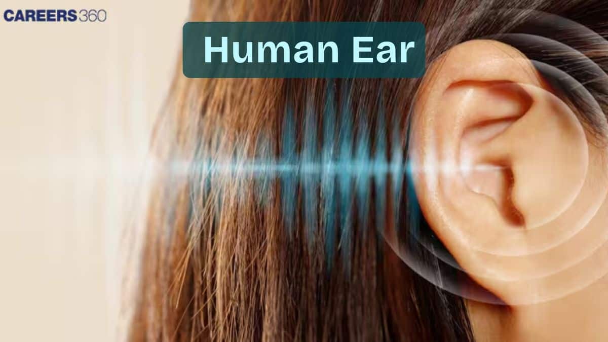
Human Ear: Structure, Function, Parts, Balance, Anatomy, Hearing
The human ear is a sensory organ responsible for hearing and balance, functioning through its outer, middle, and inner components. Sound waves are collected, amplified, and converted into electrical signals that are interpreted by the brain, while the vestibular system maintains equilibrium. This guide covers the ear’s structure, functions, diagrams, mechanism of hearing, and NEET-focused MCQs.
This Story also Contains
- Introduction — Ear as a Sensory Organ
- Structure of the Human Ear
- External Ear
- Middle Ear
- Inner Ear
- Function of the Human Ear
- Human Ear NEET MCQs (With Answers & Explanations)
- Recommended Video on Human Ear
Introduction — Ear as a Sensory Organ
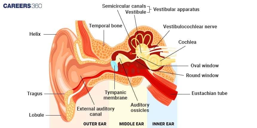
The human ear is a complex organ responsible for hearing and balance. There exist three parts of the human ear, the outer ear, which collects the sound, the middle ear, in which the collected sound gains amplification, and the inner ear, where the sound vibrations are changed into signals that reach to be read by the brain. The inner ear also helps the body maintain its balance. These structures work together for us to hear sounds and balance ourselves.
Structure of the Human Ear
The human ear consists of three main parts:
External Ear – collects sound
Middle Ear – amplifies sound
Internal Ear – hearing + balance
External Ear
The external ear is divided into the following parts:
Auricle (Pinna)
The auricle, or pinna, is a thin plate of elastic cartilage covered by a layer of skin.
Its funnel-like curves collect sound waves and transmit them to the middle ear. The lobule consists of adipose and fibrous tissues supplied with blood capillaries.
External Auditory Meatus
The external auditory meatus is a slightly curved canal supported by bone in its interior part and cartilage in the exterior part.
The canal is lined with stratified epithelium and wax glands.
Tympanic Membrane
The tympanic membrane separates the middle ear from the external ear.
This membrane receives and amplifies sound waves. Its central part is known as the umbo.
Middle Ear
The middle ear comprises the following parts:
Tympanic Cavity
The tympanic cavity is a narrow, air-filled cavity separated from the external ear by the tympanic membrane and from the inner ear by a bony wall.
It contains an auditory tube known as the eustachian tube in its anterior wall.
Eustachian Tube
The eustachian tube is a 4 cm long tube that equalizes air pressure on either side of the tympanic membrane.
It connects the tympanic cavity with the nasopharynx.
Ear Ossicles
The ear ossicles transmit sound waves from the eardrum to the middle ear. There are three ear ossicles:
Malleus: A hammer-shaped part attached to the tympanic membrane through its handle and to the incus through its head. It is the largest ear ossicle.
Incus: An anvil-shaped ear ossicle connected with the stapes.
Stapes: The smallest ossicle and the smallest bone in the human body.
Inner Ear
The inner ear comprises two main parts:
Bony Labyrinth
The bony labyrinth consists of a vestibule, three semi-circular canals, and a spirally coiled cochlea. It is filled with perilymph.
Membranous Labyrinth
The bony labyrinth surrounds the membranous labyrinth, which contains sensory receptors responsible for balance and hearing.
The membranous labyrinth is filled with endolymph and includes three semi-circular ducts, the cochlear duct, saccule, and utricle.
The sensory receptors are cristae, the organ of Corti, and ampullary maculae.
Function of the Human Ear
The human ear performs two primary functions: hearing and maintaining balance.
Hearing
The mechanism of hearing involves the following steps:
Sound waves pass through the auditory canal and reach the eardrum.
The vibrations produced pass through the tympanic membrane to the tympanic cavity.
The ear ossicles in the tympanic cavity receive the vibrations, and the stapes push the oval window in and out.
This action is transmitted to the organ of Corti, the receptor of hearing, which contains tiny hair cells that translate the vibrations into electrical impulses transmitted to the brain by sensory nerves.
Balance
The eustachian tube and the vestibular complex are crucial for maintaining balance.
The Eustachian tube equalizes air pressure in the middle ear, helping to maintain balance.
The vestibular complex contains receptors that maintain body balance.
Human Ear NEET MCQs (With Answers & Explanations)
Important questions asked in NEET from this topic are:
Structure of the human ear
Functions of the human ear
Practice Questions for NEET
Q1. The supportive skeletal structures in the human external ears and in the nose tip are examples of:
Areolar tissue
Bone
Cartilage
Ligament
Correct answer: 1) Cartilage
Explanation:
Pinna: The outer ear comprises the pinna and the external auditory meatus (canal). The pinna functions to collect air vibrations that produce sound, directing them into the external auditory meatus, which extends inward to the tympanic membrane (eardrum), where sound waves are further processed. Skeletal System: The skeletal system is a framework composed of bones and cartilage, playing a crucial role in providing structural support and facilitating movement. Among the cartilages, hyaline cartilage is the most abundant type, providing flexibility and support while reducing friction in joints.
Hence the correct answer is option 3) Cartilage.
Q2. The kind of tissue that forms the supportive structure in our pinna (external ears) is also found in:
Nails
Ear ossicles
Tip of the nose
Vertebrae
Correct answer: 1) Tip of the nose
Explanation:
There are three types of cartilage: hyaline cartilage, fibrous cartilage, and calcified cartilage. Fibrous cartilage has well-developed fibres in its matrix and can be further classified into two types fibrous cartilage which has white strong fibres, and is located in intervertebral discs and pubic symphysis, and yellow elastic fibrous cartilage with yellow fibres that provide elasticity and is located in the ear pinna and the tip of the nose.
Hence, the correct answer is option 3) Tip of the nose.
Q3. Assertion: The outer ear (external ear) is also called the auricle or pinna, your outer ear consists of ridged cartilage and skin, and it contains glands that secrete ear wax.
Reason: Its funnel-shaped canal leads to your eardrum or tympanic membrane.
If both Assertion & Reason are true and the reason is the correct explanation of the assertion, then mark A
If both Assertion & Reason are true but the reason is not the correct explanation of the assertion, then mark B
If Assertion is true statement but Reason is false, then mark C
If both Assertion and Reason are false statements, then mark D
Correct answer: 1) If both Assertion & Reason are true and the reason is the correct explanation of the assertion, then mark A
Explanation:
Both Assertion & Reason are true and the reason is the correct explanation of the assertion. Your outer ear is the part of your ear that’s visible. It’s what most people mean when they say “ear.” Also called the auricle or pinna, your outer ear consists of ridged cartilage and skin, and it contains glands that secrete ear wax. Its funnel-shaped canal leads to your eardrum or tympanic membrane.
Hence, the correct option is 1) Both Assertion & Reason are true and the reason is the correct explanation of the assertion.
Also Read:
Recommended Video on Human Ear
Frequently Asked Questions (FAQs)
The human ear works by collecting sound waves through the outer ear, which causes the vibration. These vibrations are passed through the ossicles to the inner ear, where the cochlea converts the vibrations into electrical signals. These signals are sent to the brain via the auditory nerve, allowing us to hear.
The main functions of the ear are hearing and balance. It collects sound, amplifies it, and converts sound vibrations into electrical signals that are sent to the brain for interpretation.
The ossicles amplify sound vibrations and transmit them to the inner ear.
The cochlea converts sound vibrations into electrical signals for the brain to interpret.
The semicircular canals detect head movements and help maintain balance.
The eardrum responds to sound waves and transmits these vibrations to the ossicles.