Download Careers360 App
Structure Of A Nerve

Structure Of A Nerve

Edited By Irshad Anwar | Updated on Jul 02, 2025 07:23 PM IST

What Is A Nerve?

The nervous system is formed by the cells and tissues that, in structure, are part of a systematized network having certain grades; it is through these specialized cells that the functions of the body are coordinated and controlled through electricity transferred between them. It further coordinates the integration of sensual information and the activities of muscles, while maintaining homeostasis.

On these grounds, nerves may be considered the conduit through which organs communicate with each other. This is by information on the structure nervous system, functions of the nervous system and functions of nerves in impulse transmission. It also contains information on the various types of neurons and the functions of the neurons.

What Is A Nerve?

Nerves are surrounded by connective tissue sheaths that may conduct electrical impulses between the central nervous system and the peripheries of the body or the organs or muscles in the body system. These are communicated via pathways by which the brain and spinal cord convey information to various parts of the body. They, therefore, become very vital in sensory perception; motor control and coordination of body functions.

The Basic Function Of Nerves

At the most primitive level, the nerves are primarily used for signal transmission from sensory receptors to the central nervous system and from the CNS to muscles and glands. For example, information regarding stimuli such as touch and temperature of the surroundings is carried to the brain with the help of sensory nerves. Mixed nerves have both sensory and motor nerve fibres to provide complete intercommunication between different parts of the body.

NEET Highest Scoring Chapters & Topics
Know Most Scoring Concepts in NEET 2024 Based on Previous Year Analysis.
Know More

Types Of Nerves

  • Sensory Nerves: These are afferent nerves, which carry messages from the sensory receptors in the skin, eyes, ears, nose, and tongue through the spinal cord to the brain, which makes it possible for a person to see more light, hear more sound, and feel more touch.

  • Motor Nerves: These are efferent nerves, which carry messages from the brain to the muscles and glands in the body to command all sorts of voluntary and.

  • Mixed Nerves: Such nerves contain both sensory and motor fibers and, therefore provide two-way traffic communication between the CNS and peripheral tissues.

Anatomy Of A Nerve

The anatomy of the nerve is described below-

Structure Of A Nerve

The detailed structure is explained below:

Neuron:

The aggregation of a large number of axons together forms a bundle; this bundle is covered by several layers of connective tissue to make a nerve. Axons are long, slender projections of the neuron that carry electrical impulses. The structure of the nerve accounts for the effective transmission of signals and also protects the axon from damage.

Detailed explanation of a neuron's structure

The neuron is, in fact, the simplest unit of the nerve, which is in a literal sense carrying the electrical impulse. The major components of the neuron are pointed out in the following lines.

Cell Body—Soma: There lies the nucleus and organelles, and hence it forms the metabolic part of the neuron.

Dendrites: These are the small branches coming out and receiving the signals from other neurons and passing them to the cell body.

Axon: It is the elongated projection that conducts electrical impulses away from the cell body towards other neurons or effector cells.

Myelin Sheath

It is a layer of fat, and thus it insulates the axon. The speed of transmission of impulses thus increases. The Schwann cells produce it in the peripheral nervous system and the oligodendrocytes produce it in the central nervous system.

Nodes Of Ranvier

Gaps between the myelin sheaths. They allow the impulse to travel rapidly and continuously through a process called saltatory conduction.

Axon Terminals

The ends of the axon. Neurotransmitters then spill out into other neurons or muscle cells and communicate with them.

Supporting Structures

The supporting structures are described below-

  • Endoneurium: The very thin layer of Connective Tissue surrounding individual nerve fibres within a nerve.

  • Perineurium: It is a covering of areolar connective tissue that goes into wrapping each fascicle of nerve fibres. The two main functions are extra protection and adding strength.

  • Epineurium: It refers to the outermost connective tissue wrapping layer around the entire nerve. Its main function is to provide some structural integrity and protection.
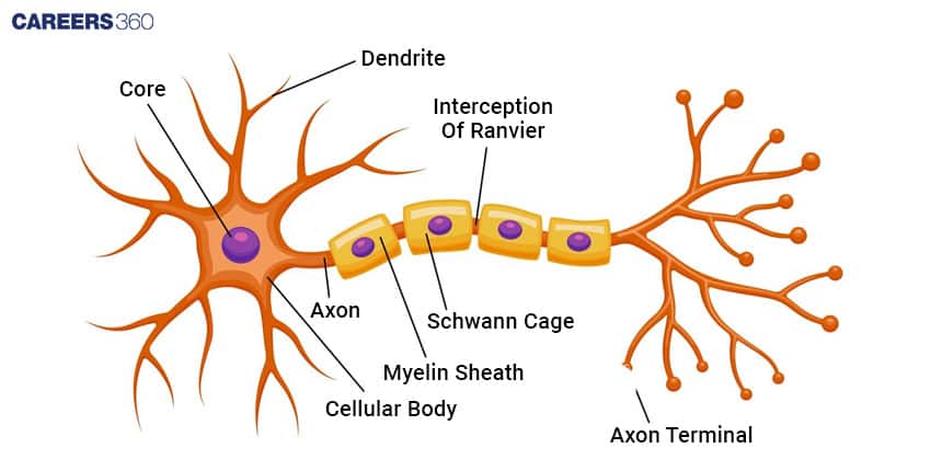
Types Of Nerve Fibers

The types of nerves are described below-

Classification Based On Diameter And Speed Of Conduction

  • Fibres (Alpha, Beta, Gamma, Delta): These are the myelinated nerve fibres of varying diameters and thus have varying conduction velocities. They are ascribed to conduct motor commands and sensory information at a fast pace.

  • B Fibers: These are the myelinated fibres of intermediate diameters and conduction velocities. They are responsible for autonomous functions.

  • C Fibers: Unmyelinated ones of small diameters with slow conduction velocities that transmit painful sensations and temperature.

Recommended video on "Structure Of A Nerve"


Frequently Asked Questions (FAQs)

1. What is the basic structure of a nerve?

The general anatomy of a nerve includes a neuron, myelin sheath, endoneurium, perineurium and epineurium.

2. What is the basic structure of a nerve?
A nerve is composed of bundles of nerve fibers (axons) wrapped in connective tissue layers. The outermost layer is called the epineurium, which surrounds multiple fascicles. Each fascicle is enclosed by the perineurium, and within fascicles, individual axons are surrounded by the endoneurium.
3. How does the myelin sheath affect nerve impulse transmission?

The myelination of axons with the myelin sheath increases its velocity in conducting an impulse of a nerve through saltatory conduction.

4. What are the different types of nerve fibres and their functions?

Nerve fibres are classified as A, B, and C fibres that vary in terms of diameter, amount of myelination, and speed of conduction as well as functions.

5. Can nerves regenerate after injury?

Yes, nerves of the PNS are regenerative with the assistance of the Schwann cells in offering locations with which the fibres can grow. There is very little, if any, capacity in the CNS for the regrowth to take place in the regeneration process.

6. What are some common nerve disorders and their symptoms?

Probably the most known nerve disorders include neuropathy, multiple sclerosis, and ALS or Lou Gehrig's disease. Each of these is very different etiologies with different symptoms associated with it.

7. How does the structure of a nerve ending differ from the main nerve fiber?
Nerve endings, or synaptic terminals, are specialized structures at the end of axons. Unlike the main nerve fiber, they lack a myelin sheath and instead contain synaptic vesicles filled with neurotransmitters. These structures are adapted for the release of neurotransmitters to communicate with target cells.
8. How does the structure of a nerve fiber support its ability to conduct electricity?
The nerve fiber's structure supports electrical conduction through its cylindrical shape, which allows for the flow of ions. The myelin sheath acts as an insulator, preventing current leakage. The nodes of Ranvier, where the axon is exposed, allow for the regeneration of the action potential, enabling efficient signal propagation.
9. What is the significance of the endoneurium in nerve structure?
The endoneurium is the innermost layer of connective tissue in a nerve. It surrounds individual nerve fibers (axons) and provides support and protection. The endoneurium also contains small blood vessels that supply nutrients to the nerve fibers.
10. What is the role of glial cells in nerve structure?
Glial cells, such as Schwann cells in the peripheral nervous system and oligodendrocytes in the central nervous system, play crucial roles in nerve structure. They form the myelin sheath, provide support and nutrition to neurons, and help maintain the appropriate environment for nerve function.
11. What is the role of Schwann cells in nerve structure?
Schwann cells play a crucial role in the peripheral nervous system by forming the myelin sheath around axons. They wrap around the axon multiple times, creating insulating layers that enhance the speed of nerve impulse transmission through saltatory conduction.
12. What is the function of the epineurium in nerve structure?
The epineurium is the outermost layer of connective tissue surrounding a nerve. It provides protection and support to the nerve, helps maintain its shape, and contains blood vessels that supply nutrients to the nerve fibers.
13. What is the role of the perineurium in nerve structure?
The perineurium is a layer of connective tissue that surrounds individual fascicles within a nerve. It provides a protective barrier, helps maintain the internal environment of the fascicle, and contributes to the nerve's mechanical strength.
14. How does the nodes of Ranvier contribute to nerve function?
Nodes of Ranvier are gaps in the myelin sheath where the axon membrane is exposed. They allow for the regeneration of action potentials, enabling saltatory conduction. This "jumping" of the electrical signal from node to node significantly increases the speed of nerve impulse transmission.
15. What is the significance of the myelin sheath in nerve structure?
The myelin sheath acts as an electrical insulator, preventing the loss of electrical current from the axon. This insulation allows for faster and more efficient transmission of nerve impulses through saltatory conduction, where the signal "jumps" from one node of Ranvier to the next.
16. What is the difference between myelinated and unmyelinated nerve fibers?
Myelinated nerve fibers have a myelin sheath surrounding the axon, formed by Schwann cells in the peripheral nervous system. Unmyelinated fibers lack this sheath. Myelinated fibers conduct impulses faster due to saltatory conduction, while unmyelinated fibers conduct impulses more slowly but continuously.
17. How does the structure of sensory nerves differ from motor nerves?
Sensory and motor nerves have similar basic structures, but they differ in the direction of signal transmission. Sensory nerves carry signals from sensory receptors to the central nervous system, while motor nerves carry signals from the central nervous system to muscles or glands. The arrangement and types of nerve fibers within these nerves may also differ.
18. How does the structure of a nerve fiber relate to its function?
The structure of a nerve fiber is specialized for rapid transmission of electrical signals. The axon is surrounded by a myelin sheath, which acts as an insulator and allows for saltatory conduction. This structure enables faster and more efficient propagation of action potentials along the nerve fiber.
19. Why are some nerves thicker than others?
Nerves vary in thickness due to the number and size of nerve fibers they contain. Thicker nerves generally have more nerve fibers or larger diameter axons. The thickness can also be influenced by the amount of myelin and connective tissue present.
20. How does the structure of a nerve fiber change after injury?
After injury, the nerve fiber undergoes a process called Wallerian degeneration. The part of the axon distal to the injury site degenerates, while the proximal part may start to regenerate. Schwann cells play a crucial role in guiding the regenerating axon and reforming the myelin sheath.
21. How does the structure of a nerve trunk differ from that of a single nerve fiber?
A nerve trunk is a bundle of many individual nerve fibers (axons) wrapped together by connective tissue. It contains multiple fascicles, each surrounded by perineurium, and the entire trunk is enclosed by epineurium. In contrast, a single nerve fiber consists of one axon, its myelin sheath (if present), and surrounding endoneurium.
22. What is the purpose of the axoplasm in nerve structure?
The axoplasm is the cytoplasm within the axon of a nerve fiber. It contains various organelles, cytoskeletal elements (like microtubules and neurofilaments), and proteins necessary for axonal transport. The axoplasm plays a crucial role in maintaining the structure of the axon and facilitating the movement of materials along its length.
23. What is the significance of the axon initial segment in nerve structure?
The axon initial segment is the specialized region of the axon closest to the cell body. It has a high concentration of voltage-gated sodium channels and is typically the site where action potentials are initiated. This structure is crucial for integrating incoming signals and determining whether the neuron will fire an action potential.
24. How does the structure of a nerve fiber change with age?
As nerves age, several structural changes can occur. The myelin sheath may become thinner or develop abnormalities. The number of nerve fibers may decrease, and the remaining fibers might shrink in diameter. These changes can lead to slower nerve conduction and reduced sensory and motor function in older individuals.
25. What is the role of the axon terminal in nerve structure and function?
The axon terminal is the specialized end of a nerve fiber where it forms a synapse with another cell. Its structure is adapted for neurotransmitter release, containing synaptic vesicles filled with neurotransmitters, mitochondria for energy production, and a high concentration of calcium channels. This structure enables the conversion of electrical signals into chemical signals for intercellular communication.
26. How does the diameter of an axon affect its function?
The diameter of an axon directly influences the speed of nerve impulse conduction. Larger diameter axons conduct impulses faster than smaller ones because they have less internal resistance to the flow of electrical current. This is why some nerves, like those controlling reflexes, have larger diameter axons.
27. What is the purpose of the axon hillock in nerve structure?
The axon hillock is the region where the axon emerges from the cell body (soma) of a neuron. It plays a crucial role in initiating action potentials. This region has a high concentration of voltage-gated sodium channels, making it the most excitable part of the neuron and the site where action potentials are typically generated.
28. How does the structure of a synapse relate to its function?
A synapse is the junction between two neurons or between a neuron and a target cell. Its structure is specialized for neurotransmission. The presynaptic terminal contains synaptic vesicles filled with neurotransmitters, while the postsynaptic membrane has receptors for these neurotransmitters. This structure allows for the chemical transmission of signals between cells.
29. What is the significance of the Ranvier constriction in nerve structure?
The Ranvier constriction, or node of Ranvier, is a gap in the myelin sheath where the axon is exposed. These nodes are crucial for saltatory conduction, allowing the action potential to "jump" from node to node. This structure significantly increases the speed of nerve impulse transmission in myelinated fibers.
30. How does the structure of a nerve fiber change as it approaches its target?
As a nerve fiber approaches its target, it typically branches into smaller fibers. The myelin sheath becomes thinner and eventually disappears near the nerve ending. The axon terminal may expand into a synaptic bulb containing synaptic vesicles, adapting the structure for neurotransmitter release.
31. What is the role of ion channels in nerve structure and function?
Ion channels are protein structures embedded in the nerve cell membrane. They play a crucial role in generating and propagating action potentials by allowing specific ions (like sodium and potassium) to move in and out of the cell. The distribution and types of ion channels along the nerve fiber are essential for its electrical properties.
32. How does the structure of a neuromuscular junction differ from a typical synapse?
A neuromuscular junction is a specialized synapse between a motor neuron and a muscle fiber. While it shares basic features with other synapses, it has a larger synaptic cleft and more extensive postsynaptic folding. The postsynaptic membrane contains a high concentration of acetylcholine receptors, adapting it for rapid and reliable neuromuscular transmission.
33. How does the structure of a nerve fiber support bidirectional transport?
The structure of a nerve fiber supports bidirectional transport through its cytoskeletal elements, particularly microtubules. These structures act as "tracks" along which motor proteins can move cargo in both directions. Anterograde transport (toward the axon terminal) is facilitated by kinesin proteins, while retrograde transport (toward the cell body) is mediated by dynein proteins.
34. What is the role of the myelin sheath in energy conservation within a nerve?
The myelin sheath conserves energy in nerve transmission by allowing saltatory conduction. This reduces the number of ion channels that need to be activated along the axon, minimizing the energy required to restore ion gradients after an action potential. As a result, myelinated fibers can transmit signals faster and more efficiently than unmyelinated fibers.
35. What is the significance of the Schmidt-Lanterman incisures in nerve structure?
Schmidt-Lanterman incisures are funnel-shaped interruptions in the myelin sheath of peripheral nerves. They provide a path for the movement of materials between the Schwann cell body and the inner layers of myelin. These structures are thought to play a role in the maintenance and nutrition of the myelin sheath.
36. How does the structure of a nerve fiber support its ability to regenerate?
The structure of a nerve fiber supports regeneration through several features. The cell body contains the genetic material and organelles necessary for producing new axonal components. The endoneurium provides a guide for regenerating axons. Schwann cells can dedifferentiate and proliferate to support regrowth and remyelination of the regenerating axon.
37. How does the structure of a nerve fiber support the process of neurotransmitter release?
The structure of the axon terminal supports neurotransmitter release through several features. It contains numerous synaptic vesicles filled with neurotransmitters, a high concentration of voltage-gated calcium channels, and proteins necessary for vesicle fusion with the membrane. The presynaptic membrane has specialized active zones where vesicles dock and release their contents into the synaptic cleft.
38. What is the significance of the Schwann cell nucleus in nerve structure?
The Schwann cell nucleus is crucial for the production of myelin and the maintenance of the myelin sheath in peripheral nerves. It contains the genetic material necessary for synthesizing myelin proteins and lipids. The position of the nucleus within the Schwann cell also creates the characteristic appearance of internodal regions in myelinated fibers.
39. How does the structure of a nerve fiber change during demyelinating diseases?
In demyelinating diseases, the myelin sheath is damaged or destroyed. This can lead to the exposure of large portions of the axon, disruption of saltatory conduction, and potential axonal degeneration. Schwann cells may attempt to remyelinate, but the new myelin sheaths are often thinner and less effective, leading to impaired nerve function.
40. What is the role of the paranodal region in nerve structure?
The paranodal region is the area on either side of a node of Ranvier where the myelin sheath terminates. It contains specialized junctions between the axon and myelin sheath that help to separate the electrical activity at the node from the internodal regions. This structure is crucial for maintaining the efficiency of saltatory conduction.
41. How does the structure of a nerve fiber support the process of axonal transport?
The structure of a nerve fiber supports axonal transport through its cytoskeletal elements, particularly microtubules and neurofilaments. These structures provide "tracks" along which motor proteins can move cargo. The elongated shape of the axon necessitates this transport system to move materials between the cell body and the axon terminal.
42. What is the significance of the basal lamina in nerve structure?
The basal lamina is a thin layer of extracellular matrix that surrounds Schwann cells and their associated axons in peripheral nerves. It plays a crucial role in nerve regeneration by providing a scaffold along which regenerating axons can grow. It also helps maintain the structural integrity of the nerve fiber and may play a role in cell-cell signaling.
43. How does the structure of a nerve fiber change during Wallerian degeneration?
During Wallerian degeneration, the portion of the axon distal to an injury site degenerates. The myelin sheath breaks down, and Schwann cells dedifferentiate and proliferate. Macrophages infiltrate the area to clear debris. This process prepares the environment for potential axon regeneration, with Schwann cells forming bands of Büngner to guide regrowth.
44. What is the role of the axolemma in nerve structure and function?
The axolemma is the cell membrane of the axon. It contains ion channels and pumps crucial for generating and propagating action potentials. At the nodes of Ranvier, the axolemma has a high concentration of voltage-gated sodium channels. The axolemma also plays a role in axon-glia interactions and is involved in the process of myelination.
45. How does the structure of a nerve fiber support its ability to conduct signals in only one direction?
While the structure of a nerve fiber allows for bidirectional electrical conduction, functional unidirectionality is achieved through the arrangement of synapses. The presynaptic terminal is specialized for neurotransmitter release, while the postsynaptic membrane contains receptors. This asymmetrical structure ensures that signals typically flow from the axon terminal to the target cell, not vice versa.
46. What is the significance of the internode length in nerve structure?
The internode length, which is the distance between two nodes of Ranvier, is a critical factor in determining the speed of nerve conduction. Longer internodes generally result in faster conduction velocities due to increased saltatory conduction. The optimal internode length is a balance between maximizing speed and ensuring reliable regeneration of the action potential at each node.
47. How does the structure of a nerve fiber change in response to chronic compression?
Chronic compression of a nerve can lead to structural changes including thinning of the myelin sheath, reduction in axon diameter, and potential axon loss. The connective tissue layers may thicken, particularly the perineurium, as a protective response. These changes can result in altered nerve function, including reduced conduction velocity and sensitivity.
48. What is the role of mitochondria in nerve structure and function?
Mitochondria are crucial organelles in nerve fibers, particularly concentrated in areas of high energy demand such as the nodes of Ranvier and synaptic terminals. They provide the ATP necessary for maintaining ion gradients, supporting axonal transport, and fueling neurotransmitter synthesis and release. Their distribution along the axon is essential for the nerve's energy supply.
49. How does the structure of a nerve fiber support its ability to conduct multiple signals simultaneously?
A single nerve contains multiple axons, each capable of conducting signals independently. Within each axon, the all-or-nothing nature of action potentials and the refractory period prevent signal overlap. The myelin sheath and nodes of Ranvier structure in myelinated fibers allow for rapid, saltatory

Articles

Back to top