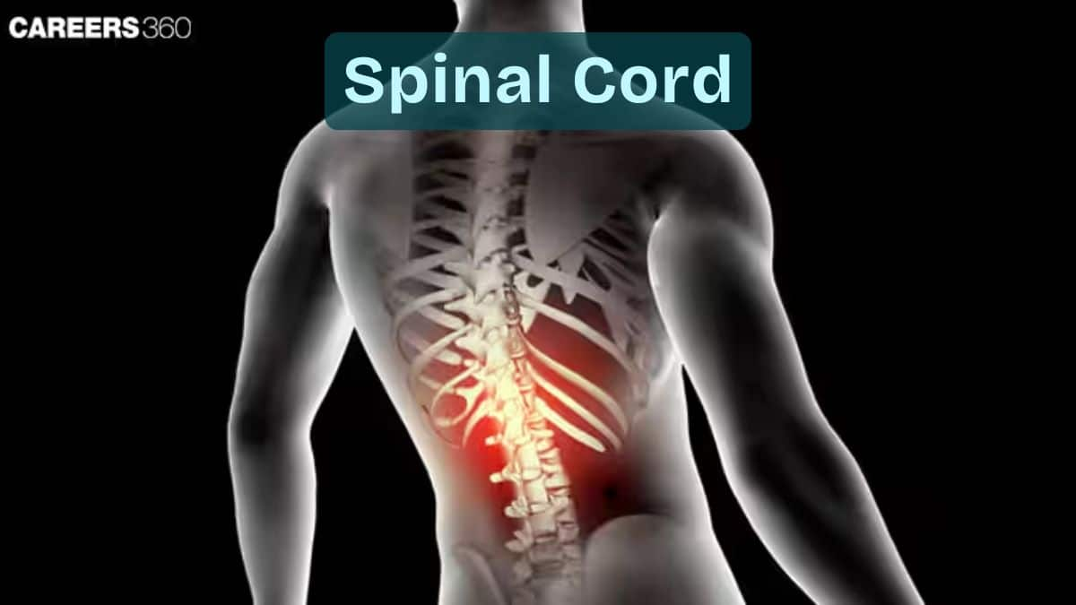
Spinal Cord: definition, meaning, function, diagram, structure
The spinal cord is the CNS pathway connecting the brain to the rest of the body, enabling movement, sensation, and reflex actions. Protected within the vertebral column, it contains 31 segments and corresponding spinal nerves that control motor and sensory functions. This guide explains spinal cord anatomy, regions, nerve functions, injuries, disorders, diagrams, NEET notes, FAQs, and MCQs.
This Story also Contains
- What Is the Spinal Cord?
- Anatomy of the Spinal Cord
- Functions of the Spinal Cord
- Spinal Cord Nerves and Their Functions
- Spinal Cord Injuries (SCI)
- Spinal Cord Disorders
- Spinal Cord NEET MCQs (With Answers & Explanations)
- Recommended Video on Spinal Cord

What Is the Spinal Cord?
It is a cylindrically shaped bundle of nerves extending from the brainstem down the vertebral column. It acts as that major pathway through which sensory and motor signals are transmitted between the brain and the rest of the body.
The spinal cord plays a crucial role in synchronising reflexes and movements, like walking and catching, with the transmission of information from the body to the brain. Housed within the protective casement of the vertebral column, it provides the basic functions that underlie simple bodily activities as well as complex motor activities, thus being indispensable to human movement, sensation, and general neurological functioning.
Anatomy of the Spinal Cord
The spinal cord is a cylindrical structure comprising nervous tissue extending from the brainstem through the vertebral column and ending near the first or second lumbar vertebra in adults. However, this may vary a bit from one individual to another.
Structure and Location
The length of the spinal cord averages approximately 45 cm (18 inches) in adults.
Its diameter varies along its length, being larger in regions where nerves controlling the limbs originate.
The total vertebral column consists of 33 vertebrae, which encase and protect the spinal cord.
There are 7 cervical, 12 thoracic, 5 lumbar, 5 sacral fused into the sacrum, and 4 coccygeal vertebrae.
Each of these vertebrae forms a part of the bony armour surrounding the fragile spinal cord, thereby protecting it from damage.
Regions of the Spinal Cord
The spinal cord is divided into:
Cervical
That part of the spinal cord in the neck region consists of 8 cervical segments (C1-C8).
Thoracic
This lies in the upper back and consists of 12 thoracic segments, T1-T12.
Lumbar
This lies in the lower back and consists of 5 lumbar segments, L1-L5.
Sacral
This is part of the pelvis and consists of 5 sacral segments, S1-S5, fused in the sacrum.
Spinal Cord Segments & Nerve Roots
The spinal cord is segmented. Each segment is related to a pair of nerve roots arising from the vertebral column.
A total of 31 segments: 8 cervical, 12 thoracic, 5 lumbar, 5 sacral, and 1 coccygeal.
Each segment gives off nerve roots that arise from the cord and emerge through openings formed by successive vertebrae.
These nerve roots then unite to form peripheral nerves that innervate regions of the body.
Functions of the Spinal Cord
A large variety of functions are carried out within the spinal cord, which includes transmitting nerve signals, controlling pathways of sensation and movement, and providing reflex actions.
Transmission of Nerve Signals
The spinal cord relays sensory information it picks up from the peripheral sensory receptors towards the brain for processing.
It receives the motor signals from the brain, triggering an outward flow that initiates muscle and other responses
Information on touch, temperature, pain, and proprioception from the body is transmitted to the brain through the spinal cord via the sensory pathways.
Coordination of Movements
They involve sensory neurons that take signals directly up the spinal cord to the brainstem and higher brain centres for perception.
The motor pathways carry commands from the brain down the spinal cord to motor neurons innervating muscles and glands throughout the body.
It's through this that the communication will allow for both voluntary movements, like walking or reaching, and involuntary actions, like regulating the heartbeat.
Reflex Actions
Reflex actions are responses that are immediate to certain stimuli, not requiring any conscious processes and directly involving the spinal cord.
The examples include protection of the body against damage, maintenance of posture, and regulation of a variety of physiological processes, all without requiring a single input from the brain.
For instance, during the patellar reflex, which is also referred to as the patellar reflex, tapping the patellar tendon initiates involuntary stretching of the leg.
Another example is the withdrawal reflex: the hand is suddenly drawn back from a hot surface.
Spinal Cord Nerves and Their Functions
Each of these segments corresponds to a specific pair of spinal nerves supplying different regions of the body. The total overview of the primary spinal nerves and their functions is given below:
Cervical Nerves (C1-C9)
C1-C4: The cervical nerves predominantly provide structures for muscles of the neck and respiration. Therefore, the nerves take part in breathing and movements of the head.
C5-C8: These nerves innervate muscles in the shoulders, arms, and hands. They play a very important role in upper limb movements and terms of sensory perception in the line of these parts.
Thoracic Nerves (T1-T12)
T1-T12: Thoracic nerves innervate the muscles and skin of the thorax and abdomen. They participate in the movements of the chest wall and the abdominal muscle activity, as well as sensitivity in those areas.
Lumbar Nerves (L1-L5)
L1-L5: Lumbar nerves innervate the lower back, buttocks, thighs, legs, and feet. They are responsible for locomotion and sensibilities in these fields, such as walking, standing, and balance.
Sacral Nerves (S1-S5)
S1-S5: These nerves are responsible for the innervation of the pelvis, genitals, buttocks and lower limbs and orchestrate acts like bowel and bladder activity, sexual function, movement and sensibility of the lower extremity.
Coccygeal Nerves (Co1)
Coccygeal Nerve (Co1): The coccygeal nerve is the smallest spinal nerve, and it innervates a small area of skin over the coccyx (tailbone).
Spinal Cord Injuries (SCI)
Trauma or disease can cause spinal cord injuries (SCI), which are divided into two major types: complete and incomplete injuries.
Complete vs Incomplete Injuries
Complete injuries: These occur with a total loss of sensation and motor function below the level of injury. The most common cause is cutting or serious damage to the spinal cord.
Incomplete Injuries: It preserves partial communication between the brain and parts of the body below the site of injury. Sensation and motor function may then be partially preserved, depending on the extent of damage to the spinal cord.
Causes of Spinal Cord Injuries
The spinal cord is prone to many disorders that either diminish its blood flow or harm it directly.
Trauma: This can be caused by everyday events, including road traffic accidents, falls, sporting injuries, and acts of violence.
Disease: All kinds of pathologies may cause spinal cord lesions, such as Tumors, infections, meningitis, and degenerative disorders, especially spinal stenosis.
Spinal Cord Disorders
The major spinal cord disorders are:
Multiple Sclerosis
A case of autoimmune neurologic disorder wherein the immune system acts against the protective myelin sheath covering nerve fibres in the brain and spinal cord. It causes problems in communication between the brain and the rest of the body.
Amyotrophic Lateral Sclerosis (ALS)
Lou Gehrig's disease is a neurodegenerative process characterized by progressive destruction of nerve cells in the brain and spinal cord. These nerve cells control voluntary muscle movement. Hence, their loss leads to the loss of muscle control, consequently resulting in paralysis.
Spina Bifida
This refers to a congenital condition whereby, before birth, there is an incomplete development of the spinal cord and its covering structures. It generates an extremely vast array of different physical and intellectual disabilities based on severity.
Spinal Cord NEET MCQs (With Answers & Explanations)
Important questions asked in NEET from this topic are:
Anatomy of spinal cord
Functions of spinal cord
Disorders related to spinal cord
Practice Questions for NEET
Q1. The spinal cord extends from the foramen magnum at the base of the skull to the L1/L2 vertebra where it terminates as the
Conus medullaris (medullary cone)
Coccygeal vertebra
Epiduramater
Choroid plexus
Correct answer: 1) Conus medullaris (medullary cone)
Explanation:
The spinal cord, an essential component of the central nervous system, originates from the foramen magnum, a significant aperture situated at the skull's base, and extends down to the L1/L2 vertebral levels. It does not stretch throughout the vertebral column's entirety, but rather concludes at this point, transforming into the conus medullaris. Following the conus medullaris, the cauda equina, a collection of spinal nerve fibres, descends within the vertebral canal.
Hence, the correct answer is option 1. Conus medullaris (medullary cone).
Q2. The number of pairs of thoracic spinal nerves is
31
12
5
8
Correct answer: 2) 12
Explanation:
There are 8 pairs of cervical, 12 thoracic, 5 lumbar, 5 sacral, and 1 coccygeal pair of spinal nerves (a total of 31 pairs).
Hence, the correct answer is option 2) 12.
Q3. Identify the following statements.
1) The neural canal is the channel for the spinal cord to pass through.
2) The gray matter extends from the brain to the inside of the spinal cord and white matter covers the surroundings of the spinal cord.
Statement 1 is false.
Statement 2 is true.
Statements 1 and 2 are true
Statements 1 and 2 are false.
Correct answer: 3) Statements 1 and 2 are true
Explanation:
Both are true of the spinal cord:
The spinal cord is a long, cylindrical structure running from the back of the brain down to the lower part of the back and through the neural canal. It is an extremely important linkage between the central and peripheral nervous system.
The sensory neurons transmit impulses through the spinal cord. It is the portion that contains grey matter as horn-like projections and white matter surrounding it. Grey matter includes the cell bodies of neurons while white matter consists of myelinated axons connecting to other parts of the nervous system.
The spinal cord carries out the reception of sensory input and coordination of reflexes.
Hence the correct answer is option 3) Statements 1 and 2 are true.
Also Read:
Recommended Video on Spinal Cord
Frequently Asked Questions (FAQs)
The spinal cord's main function is the conduction of sensory information from the body towards the brain and motor commands from the brain towards the body.
Common causes of spinal cord injuries include traumatic events like motor vehicle accidents, falls, sports injuries, and acts of violence. Nontraumatic events caused by diseases include spinal cord tumours, infections and degenerative disorders.
Grey matter consists mainly of neuronal cell bodies, dendrites, and unmyelinated axons of the spinal cord. White matter consists of myelinated axons that form nerve tracts connecting regions of the spinal cord and convey signals between the brain and the body.
Research is being conducted on spinal cord regeneration through various methods to enhance nerve regeneration and regain function, such as stem cell therapy, nerve grafting, and biomaterial scaffolds.
The diagnosis of spinal cord injury includes clinical evaluation for imaging tests to visualise damage to the spinal cord; neurological examinations delineate sensory and motor loss below the level of injury.
