

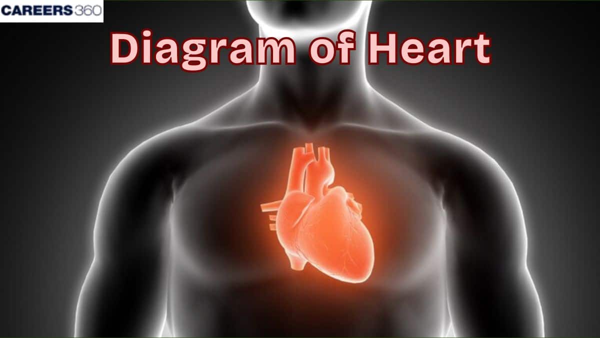
Diagram of Heart
The human heart is a muscular organ responsible for pumping oxygenated and deoxygenated blood across the body. Understanding its anatomy—chambers, valves, layers, and blood vessels—is essential for NEET Biology and Class 11/12 Circulation chapter. This guide explains the structure, functions, and clinical significance of the heart with diagrams, MCQs, FAQs, and exam-oriented notes.
This Story also Contains
- What is Heart?
- Functions of the Human Heart
- Anatomy of Human Heart
- Chambers of the Heart
- Valves of the Human Heart
- Layers of the Heart Wall
- Disorders
- Heart NEET MCQs (With Answers & Explanations)
- Recommended Video for Heart
What is Heart?
The heart is one of the principal human body organs that play a crucial role in the circulatory system as it is in charge of the circulation of blood in the body to facilitate aeration, supply nutrients and expulsion of metabolic wastes. Knowing the human heart is necessary for comprehending the simple vital and ruling life procedures and their importance in the clinical analysis and treatment of diseases. Appreciation of these ideas is essential, especially for individuals seeking to become healthcare professionals as they determine how the patient’s clinical signs present, how to handle the patients, and how to manage the patients’ conditions appropriately.
Functions of the Human Heart
The functions of the human heart are discussed below:
The force and rate of the contractions of the heart helps to avoid inadequate blood flow and hence maintains blood pressure in the body..
The circulatory system transports material such as glucose and amino acids in their developed forms to cells and embraces products such as carbon dioxide and urea.
It also plays the role of an endocrine gland because it has the capability of secreting hormones that control other activities in the body. Atrial Natriuretic Factor (ANF) is released from the atria of the heart when blood volume and pressure are high.
Anatomy of Human Heart
The human heart is a muscular organ located in the chest that consists of four chambers and which pumps blood to and from the body. The anatomy of the human heart is discussed below:
Location And Size
The human heart is situated in the mediastinum, more specifically in the thoracic cavity and in front of the lungs in coordination with the left side of the sternum axis.
It is situated behind the sternum and above the diaphragm.
An average human heart is as large as a fist and weighs between 250-350gm (9-12oz) in an adult.
Pericardium
The overall structure in which the heart is surrounded is called the pericardium and is made up of two elementary structures, the fibrous pericardium and the serous pericardium.
Serous pericardium is subdivided into the parietal and visceral layers.
The pericardium acts as a shield to the heart, it holds the heart in position and acts as a constraint to the heart’s ventricular filling.
Chambers of the Heart
The heart's major parts include:
Atria
Right Atrium: Gathers deoxygenated blood from the body and directs it to the right atrium of the heart through this S-shaped tube.
Left Atrium: Recovers O2-rich blood from the lungs and pumps it into the left ventricle chamber.
Ventricles
Right Ventricle: Pumps deoxygenated blood to the lungs for oxygenation increasing the level of oxygen in the bloodstream.
Left Ventricle: Kills off low-oxygenated blood in the pulmonary veins and pumps oxygenated blood to all parts of the body.
Valves of the Human Heart
There are four valves that ensure unidirectional blood flow including the tricuspid, pulmonary, mitral, and aortic ones.
Tricuspid Valve: It is located between the right atrium and right ventricle and this prevents the backflow of blood towards the atrium.
Pulmonary Valve: It is located in between the right ventricle and the pulmonary artery; its function is to stop the blood from flowing back into the right ventricle.
Mitral Valve: Located between the left atrium and left ventricle, its purpose is to avoid the backflow coming into the atrium.
Aortic Valve: It is situated between the left ventricle and aorta and it closes to prevent backflow into the ventricle.
Layers of the Heart Wall
The heart wall is composed of three distinct layers:
Epicardium: The outer layer, the outermost layer of very loose connective tissue that protects the target tissue.
Myocardium: The second layer is a middle layer that is a thicker portion of the cardiac muscle including the layers that cause the contractions of the heart.
Endocardium: They are grouped into four layers with layer one being the thinnest layer and the inner layer of the heart walls It is also found on the heart valves where it gives a smooth surface to the blood flow.
Disorders
Caring for our hearts is among the core values for the quality of life free from diseases, mishaps, and untimely deaths.
Coronary Artery Disease (CAD) is a condition that affects the blood vessels called the coronary arteries leading into the heart muscle and which get blocked thus limiting the amount of blood that is delivered to the muscle.
Heart attack happens when a section of the heart does not get an adequate amount of blood and nutrients hence becoming damaged.
Heart failure is a state in which the heart is not able to pump blood as it should, hence accumulating fluids within the body.
Heart NEET MCQs (With Answers & Explanations)
Types of questions asked from this topic are:
Anatomy of Human heart
Disorders related to heart
Practice Questions for NEET
Q1. The human heart is derived from
Ectoderm
Mesoderm
Endoderm
Both 1 and 3
Correct answer: 2) Mesoderm
Explanation:
Early embryonic mesoderm gives rise to the heart through a region known as the cardiogenic mesoderm. Initially, the heart is shaped as two separate tubes which merge to create a single tubular structure. This tube undergoes folding and dividing processes to develop into the atria and ventricles, along with other heart components. The mesoderm is the primary origin of the heart's tissue and structure during embryonic development.
Hence, the correct answer is option 2) Mesoderm.
Q2. The right aortic arch is found in
Mammals
Mammals and reptiles
Birds
Amphibians
Correct answer: 2) Mammals and reptiles
Explanation:
The aortic arch, the arch of the aorta or the transverse aortic arch is a part of the aorta between the ascending and descending aorta. The arch travels backwards so that it ultimately runs to the left of the trachea.
The right-sided aortic arch is a rare anatomical variant in which the aortic arch is on the right side rather than on the left.
Hence, the correct answer is option 2) Mammals and reptiles.
Q3. The pericardium and the pericardial fluid help in
Protecting the heart from friction, and shocks and keeping it moist
Pumping the blood
Receiving the blood from various parts of the body
None of these
Correct answer: 1) Protecting the heart from friction, and shocks and keeping it moist
Explanation:
The pericardium is a double-layered membrane surrounding the heart, consisting of an outer fibrous layer and an inner serous layer. It protects the heart, anchors it within the chest cavity, and prevents overexpansion during blood volume changes. The pericardial fluid, found between the serous layers, acts as a lubricant to reduce friction as the heart beats. This fluid also helps distribute mechanical forces evenly across the heart. Together, the pericardium and pericardial fluid ensure the heart functions efficiently within a stable and protective environment.
Hence, the correct answer is option 1) Protecting the heart from friction, and shocks and keeping it moist.
Also Read:
Recommended Video for Heart
Frequently Asked Questions (FAQs)
Coronary arteries provide the blood with rich oxygen to the muscular walls of the heart.
An ECG monitors the heart's electrical impulses to identify the irregularities of rhythm and conduction.
The heart circulates oxygen-rich blood to the body and the deoxygenated blood back to the lungs to be oxygenated.
Blood circulation to the right side of the heart is maintained through four valves, tricuspid, pulmonary, mitral and aortic which only let blood flow in one direction.
Systemic circulation means it supplies the oxygenated blood to the rest of the body and removes the deoxygenated blood and takes it back to the heart; Pulmonary circulation means; it takes the deoxygenated blood to the lungs where it will be oxygenated then transported back to the heart.
