

Thoracic Cavity: Meaning, Organs, Diagram, Functions
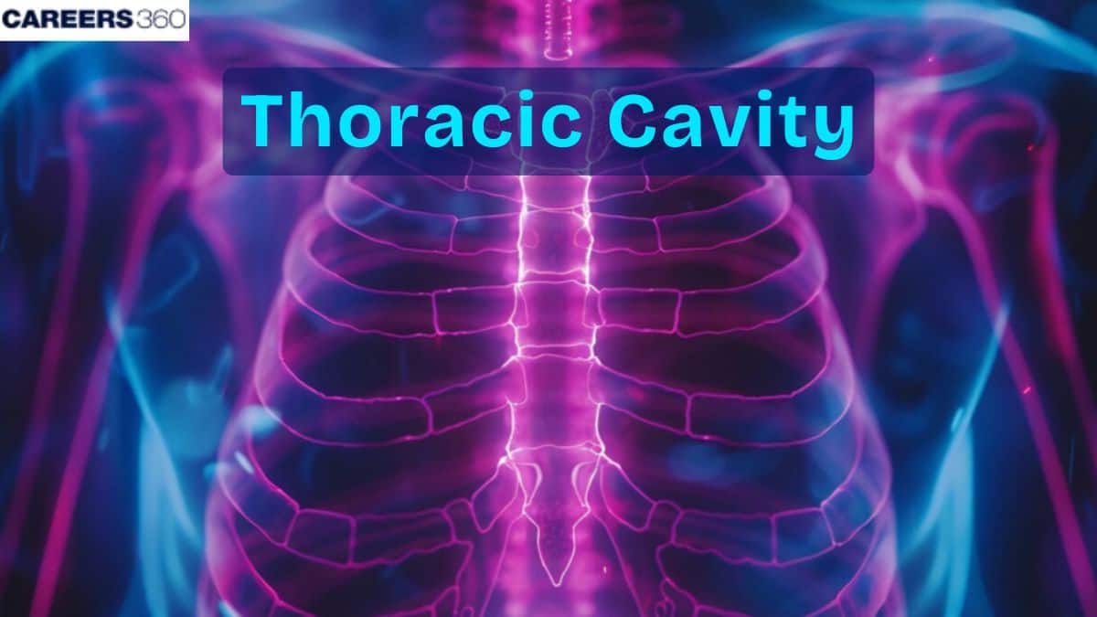
The thoracic cavity is the central chest chamber housing the heart, lungs, and major blood vessels. Protected by the ribs, sternum, diaphragm, and vertebrae, it enables essential processes like breathing and circulation. This blueprint covers its anatomy, functions, and clinical significance for NEET, nursing, and medical exams.
This Story also Contains
- What Is The Thoracic Cavity?
- Anatomy Of The Thoracic Cavity
- Functions Of The Thoracic Cavity
- Clinical Significance of the Thoracic Cavity
- Thoracic Cavity NEET MCQs (With Answers & Explanations)
- Recommended Video on "Thoracic Cavity"
What Is The Thoracic Cavity?
The thoracic cavity is a body chamber that occurs between the cervical and abdominal cavity and is bordered by the ribs, sternum, and thoracic vertebrae. It contains vital organs for example the heart and lungs, and is involved in respiration – circulation.
Knowledge of the thoracic cavity is essential for learning how these organs work and what they do, and in managing the diseases and injuries of the chest. This paper aims to focus on a detailed description of the thoracic cavity’s structure, its functions, as well as clinical considerations involving this specific cavity.
Anatomy Of The Thoracic Cavity
The anatomy of the thoracic cavity is given below-
Location and Boundaries
The thoracic cavity refers to that part of the body that is located within the trunk, specifically in the region between the neck and the abdominal cavity.
The thoracic cavity is enclosed by the thoracic wall cranially, of which the thoracic inlet forms the entrance to the thoracic cavity from the neck.
Thoracic Wall Components
Superiorly, the cavity is enclosed by the sternum whereas in the posterior aspect; it is marked by the thoracic vertebrae.
On the right angle, the rib cage supporting, and shielding the cavity is partially located.
Diaphragm
Inferiorly, it is bounded by the diaphragm, muscular mural exposes the inferior surface of the lungs and separates the thoracic cavity from the abdominal cavity.
The diaphragm acts as the floor of the thoracic cavity and is a major muscle of the respiratory system through its contraction and becoming flattened to augment the volume of the thoracic cavity during inspiration.
This muscular partition divides the thoracic cavity from the abdominal cavity and thus the integrity of this cavity is maintained.
Functions Of The Thoracic Cavity
The Thoracic Cavity plays the following important role:
Protection Of Vital Organs
The torso is made of ribs and sternum thus offering a shield to critical organs such as the heart and lungs from physical impacts from the outside world.
The thoracic vertebrae together with the vertebral columns provide extra defense as well as rigidity to the thoracic region.
Facilitation Of Breathing
Convection and circulation are mechanically enabled by the thoracic cavity for example in breathing.
The diaphragm contracts and becomes flat to make the thoracic cavity larger in the process of inhalation, while the intercostal muscles help lift the ribs.
This produces a sub atmospheric pressure within the lungs and lets air into the lungs, hence creating positive pressure.
Circulatory Functions
The left part of the thoracic cavity contains the heart, which is a part of the circulatory system since it is pumping blood in the body.
Large blood vessels specifically; the aorta and pulmonary arteries are fixed in the thoracic cavity as a way of delivering oxygenated and deoxygenated blood respectively to the heart and other parts of the body through the most effective means.
Clinical Significance of the Thoracic Cavity
The thorax can have these issues:
Thoracic Injury
Thoracic injuries, which include the ribs or any blunt force, pose a risk to the organs within the thoracic cavity which include the lungs and heart. Broken ribs can be painful and hinder the ability to breathe while severe injuries may cause internal bleeding or affect their breathing.
Respiratory Impairments
Reduced thoracic expansion limits the lungs’ ability to fully inflate, which decreases oxygen intake and compromises overall respiratory efficiency, especially during physical stress.
Diaphragm Disorders
Impaired diaphragm contraction lowers lung capacity and leads to shallow, labored breathing, making it difficult to maintain adequate ventilation and increasing fatigue.
Cardiovascular Impact
Trauma to major vessels can disrupt normal blood flow and impair organ perfusion, potentially causing rapid blood loss, hemodynamic instability, and life-threatening complications.
Thoracic Cavity NEET MCQs (With Answers & Explanations)
Important topics for NEET are:
Functions of thoracic cavity
Clinical significance of thoracic cavity
Practice Questions for NEET
Q1. Which of the following statements about the pleural cavity is incorrect?
It is a potential space between the visceral and parietal pleura.
It is filled with pleural fluid.
It helps reduce friction during breathing.
It is located within the mediastinum.
Correct answer: 4) It is located within the mediastinum.
Explanation:
The mediastinum does not contain the pleural cavity. The heart, major vessels, esophagus, and trachea are all located in the mediastinum, which is the thoracic cavity's core compartment. On the other hand, the visceral and parietal pleura, two layers of serous membrane that encircle the lungs, may be separated by the pleural cavity. Pleural fluid, which fills the pleural cavity, serves to lessen friction while breathing and enables the lungs to expand and contract without resistance.
Hence, the correct answer is option 4) It is located within the mediastinum.
Q2. Oblique fissure is present in
Left lung
Right lung
Both
None
Correct answer: 3) Both
Explanation:
The right lung consists of three lobes—superior, middle, and inferior—separated by two fissures: the horizontal fissure (between the superior and middle lobes) and the oblique fissure (between the middle and inferior lobes). In contrast, the left lung has only two lobes—superior and inferior—divided by a single oblique fissure. The left lung is slightly smaller than the right lung due to the space occupied by the heart, and it lacks the middle lobe that is present in the right lung.
Hence, the correct answer is option 3) Both.
Q3. Where are lungs situated in the human body ?
Abdominal cavity
Thoracic cavity
Coelomic cavity
Pleural cavity
Correct answer: 2) Thoracic cavity
Explanation:
The lungs are situated in an air-tight chamber called the thoracic chamber. The thoracic chamber is formed dorsally by the vertebral column, ventrally by the sternum, laterally by the ribs, and on the lower side by the dome-shaped diaphragm.
Hence, the correct answer is option 2) Thoracic cavity
Also Read:
Recommended Video on "Thoracic Cavity"
Frequently Asked Questions (FAQs)
The thoracic cavity protects such important organs, helps in the process of breathing and shrinking of the lungs as well as contributing to circulatory processes due to the housing of the heart and the major veins.
The diaphragm mainly involves the process of breathing by contracting and flattens, expanding the volume of the thoracic cavity during the inhalation process to pull in air into the lungs through the negative pressure system.
The thoracic cavity is investigated medicinally through clinical assessment, Chest X-ray, CT scan, MRI and Ultrasound. Bronchoscopy, thoracentesis and pulmonary function tests are other also used.
Pneumonia, pleuritis, lung cancer, pneumothorax and a coalition of lung, and rib fractures are some of the diseases that arise from the thoracic cavity. Some of the disorders are atelectasis and chronic obstructive pulmonary disease (COPD) affect thoracic health.
The thoracic cavity holds the heart and lungs, food pipe, windpipe and the major blood vessels including the aorta and pulmonary arteries.
