

Food Pipe/ oesophagus: Meaning, Parts, Anatomy, Function, Conditions
The oesophagus (food pipe) is a hollow, muscular tube that transports food from the pharynx to the stomach by peristaltic movements. It forms an essential part of the upper gastrointestinal tract and lies behind the trachea. This guide explains the structure, layers, functions, disorders, diagnostic techniques, and NEET-focused questions on the oesophagus.
This Story also Contains
- What Is the Oesophagus (Food Pipe)?
- Layers of the Oesophagus
- Functions of the Oesophagus
- Disorders of the Oesophagus
- Diagnostic Techniques
- Prevention and Care of the Oesophagus
- Oesophagus NEET MCQs (With Answers & Explanations)
- Recommended video for "Oesophagus"
What Is the Oesophagus (Food Pipe)?
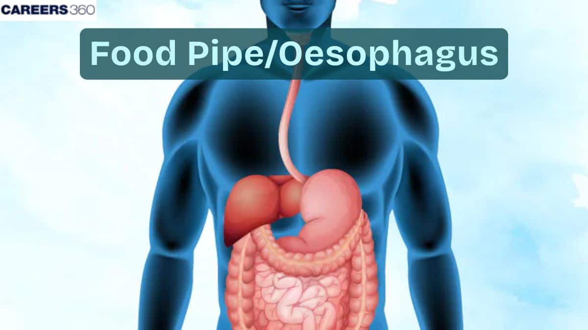
The oesophagus is a hollow, muscular tube lined internally by a mucous membrane. It is also referred to as the food pipe. That body organ is an important component of our human digestive system, passing food from the throat down towards the stomach and eventually leading to the digestive process. The oesophagus starts from the lower end of the pharynx and descends to the level of the stomach. It forms part of the upper gastrointestinal tract. It lies behind the trachea.
Layers of the Oesophagus
The oesophagus consists of four well-defined layers:
Mucosa
The innermost layer.
Secretes mucus, which helps in lubrication during the passage of food.
Protects the oesophagus from friction caused by abrasive particles.
Submucosa
Directly under the mucosa is the submucosa.
Consists of blood vessels and nerves.
Consists of mucous glands that produce mucus.
Supports the mucosa and, in return, it gives the blood supply to the mucosa.
Muscularis
Two kinds of muscles: circular and longitudinal.
Both these combine to give peristaltic movements
Moves the ingested food down the oesophagus.
Adventitia
The outermost layer is the adventitia, composed of connective tissue.
Anchors the oesophagus to surrounding structures in the neck and chest.
Functions of the Oesophagus
The oesophagus performs important functions, which include:
Transport of Food
The oesophagus is concerned mainly with the passage of food and liquids from the mouth down to the stomach.
This passage is brought about through rhythmic peristalsis.
Protection
The oesophagus protects the digestion system from the materials that cause damage and pain during swallowing.
Protects oesophagus from refluxed stomach contents
Organised Transport
The oesophagus transports all materials from the throat to the stomach
Ensures one-way passage of the materials
Disorders of the Oesophagus
The oesophagus can suffer from many diseases, but the most severe of all is oesophagal cancer.
Oesophageal Cancer
Oesophageal cancer is a malignant process that develops from the tissue of the oesophagus.
Symptoms: Difficulty swallowing, weight loss, chest pain and coughing.
Treatment: Surgical methods, radiotherapy, chemotherapy.
GERD (Gastro-oesophageal Reflux Disease)
GERD is a chronic condition where there is backflow of stomach acid to the oesophagus.
It causes irritation and consequently discomfort.
Achalasia
Achalasia is a rare disorder wherein there is difficulty in passing food from the oesophagus into the stomach
Leads in trouble swallowing food, among many other symptoms.
Esophagitis
Esophagitis is an inflammation of the oesophagus.
Causes include acid reflux, infections, and even medications for some diseases and disorders.
Diagnostic Techniques
Several diagnostic modalities available in diagnosing oesophageal disorders include the following:
Endoscopy
A long, flexible tube with a camera on the end will then be passed into the oesophagus.
It allows one to visualize the oesophagus for any abnormalities.
Barium Swallow Test
Patients swallow a barium solution.
X-ray shows the form and condition of the oesophagus.
Manometry
Manometry measures muscular contractions of the oesophagus
Detects abnormal movements or functions.
Prevention and Care of the Oesophagus
A few practices need to be followed to keep the oesophagus healthy:
Healthy Habits
- Avoiding smoking and unhealthy dietary habits.
- Maintaining a healthy weight can help avoid disorders of the oesophagus.
Screenings
- Regular follow-up medical checkups, with early diagnosis, are very important for the prevention as well as treatment of oesophagal diseases.
Medical Interventions
- There are ample treatment options available to the patient, ranging from medicines and lifestyle changes; often, even surgery.
Oesophagus NEET MCQs (With Answers & Explanations)
Important questions asked in NEET from this topic are:
Layers of the oesophagus
Disorders of the oesophagus
Practice Questions for NEET
Q1. The outer wall of the esophagus is not lined by the serous membrane. Instead, it contains
Adventitia externa
Lamina propria
Adventitia interna
Muscularis tunica
Correct answer: 1) Adventitia externa
Explanation:
As we have learned in Serosa: It is present only in the region of the alimentary canal within the abdominal cavity. Instead of serosa, the mouth, pharynx, and oesophagus have a dense sheath of collagen fibers called the adventitia. The outer wall of the oesophagus is not lined by a serous membrane, The outer wall is seen in the form of an irregular coat of yellow elastic dense fibrous connective tissue called adventitia externa or tunica adventitia.
Hence, the correct answer is option 1) Adventitia externa.
Q2. Which of the following structures prevents the entry of food particles into the trachea during swallowing?
Glottis
Uvula
Epiglottis
Both a & c
Correct answer: 3) Epiglottis
Explanation:
The Glottis is the opening of the pharynx into the trachea and is always open during the process of respiration. The uvula is a soft muscular structure preventing the entry of food into the nasal cavity during swallowing. The Glottis is covered by a flap-like structure called epiglottis which helps prevent the food particles from moving into the trachea during swallowing instead of the food pipe.
Hence the correct answer is option 3) Epiglottis.
Q3. Heartburn or gastroesophageal reflux disease is caused when
The oesophagus is open during swallowing
Movement of food from the pharynx into the esophagus is altered
The gastro-esophageal sphincter does not completely close
None of the above
Correct answer: 3) The gastro-esophageal sphincter does not completely close
Explanation:
When the gastro esophageal- sphincter does not completely close, the stomach’s contents can reflux (that is, back up into the oesophagus), causing heartburn or gastroesophageal reflux disease (GERD).
Hence, the correct answer is option 3) the gastro-esophageal sphincter does not completely close.
Also Read:
Recommended video for "Oesophagus"
Frequently Asked Questions (FAQs)
Difficulty in swallowing, weight loss, chest pain, and persistent coughing are some of the symptoms of oesophageal cancer.
Have a healthy diet, do not smoke, consume wine in moderation only, and see your doctor often for check-ups.
Tests which have been developed to investigate the disorders of the oesophagus include endoscopy, the barium swallow, and manometry.
Food and liquids move from the mouth to the stomach through the oesophagus by peristaltic movements.
Peristalsis is rhythmic contractions of the oesophageal muscles that move food downward toward the stomach.
