X-Rays - Properties, Definition, Wavelength, Types, Uses, FAQs

X-Rays - Properties, Definition, Wavelength, Types, Uses, FAQs

Team Careers360Updated on 02 Jul 2025, 05:07 PM IST

X-rays are a kind of radio waves (electromagnetic radiation), similar to visible light. Unlike light, still, x-rays have higher energy and can move or pass through most bodies, as well as the object. Medical x-rays are used to produce images of tissues and structures inside the object or body. In this article, we will discuss, who is invented or discover X-ray? What is X-ray? What is X-ray wavelength? What is the meaning of X-ray? How are X-ray produced? What is frequency of X-ray? What are uses, application and properties/characteristics of X-ray? What are the types of X-rays? So let’s see,

This Story also Contains

  1. What are X-rays?/ what is x ray?
  2. Properties of X-Rays/ properties of x rays
  3. What are the Characteristics of X Rays?
  4. x rays uses /Applications of X Rays
X-Rays - Properties, Definition, Wavelength, Types, Uses, FAQs
X-Rays

X-ray

What are X-rays?/ what is x ray?

Definition: X-Ray is also known as the “Roentgen radiation”. It is a radio wave (electromagnetic radiation) with an energy which ranges from 124 eV to 124 keV. where this energy can be put in the form of J (Joules). Still, a wave with this more energy can easily transfer from non-opaque (transparent) to non-transparent (opaque) objects. The meaning of X-ray in hindi “क्ष-किरण” “ऍक्स-किरण”.

Who invented or discovered X-ray?

On 8 November, 1895, X-rays were discovered by a German Physicist called “Wilhelm Conrad Röentgen”. X-rays were discovered fortuitously by German scientist Roentgen in 1895. In 1901, Roentgen was awarded for his great add this regard. German physicist Wilhelm Röntgen is usually credited for the invention of X-Rays in 1895 because he was the primary to extensively examine them, though he's not thought to be the primary to possess seen and perceived their effects. They were found emanating from Crookes tubes, experimental discharge tubes invented around 1875, by scientists looking into the cathode rays, which are energetic electron beams that were first formed within the tubes. X-rays are highly penetrating electromagnetic wave and have proved to be a really powerful tool to review the crystal structure, in material research, within the radiography of metals and within the field of medical sciences.

Also read -

What is the wavelength of X-ray?/ What is x ray wavelength?

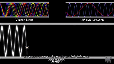
X-ray is an electromagnetic wave with very small wavelength, and really greater energy. X rays have a frequency starting from 30 petahertz to 30 exahertz.

The wavelength of X-rays is smaller than the Ultraviolet rays, and greater than Gamma rays.

So, what's the wavelength of x rays?

X Rays have a wavelength ranging from 0.01 to 10nm.

How X-rays are produced?/ how are x rays produced?/ x ray production
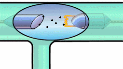
production of X ray

Roentgen discovered that when X-rays are skilled arms and hands or the other part.

Experimentally, x rays are produced from a high energetic electron beam. When the beam is incident on a target, x-rays are formed. The target should have high melting point and high atomic mass.

How do X-rays work?

1642676764237

They are generated when high-velocity electrons hook the metal plates, consequently giving the energy because the X-Rays and themselves are absorbed by the metal plate.

The X-Ray beam travels via the air and comes in touch with the body tissues, and generates a picture on a metal film.
1642676762673

Soft tissue such as organs and skin cannot soak up (absorb) the high-energy rays, and consequently the beam passes via them.

Dense materials inside our bodies, like bones, soak up the radiation.

Commonly Asked Questions

Q: What is synchrotron radiation and how is it related to X-rays?
A:
Synchrotron radiation is electromagnetic radiation emitted when charged particles, usually electrons, are accelerated in a curved path. Modern synchrotron facilities produce intense, tunable X-rays used in various scientific applications, from materials science to structural biology.
Q: What is X-ray photoelectron spectroscopy (XPS) and how is it used?
A:
XPS is a surface-sensitive technique that uses X-rays to eject electrons from a material's surface. By analyzing the energy of these ejected electrons, scientists can determine the elemental composition and chemical state of the surface atoms, with applications in materials science and catalysis research.
Q: What is X-ray microscopy and how does it complement other microscopy techniques?
A:
X-ray microscopy uses X-rays to create high-resolution images of samples, offering a resolution between that of optical and electron microscopy. It can penetrate thicker samples than electron microscopy and provide elemental contrast, making it valuable for studying biological specimens and materials science samples.
Q: What is X-ray lithography and how is it used in semiconductor manufacturing?
A:
X-ray lithography is a technique used to create extremely small patterns on semiconductor wafers. It uses X-rays to transfer a geometric pattern from a mask to a light-sensitive chemical photoresist on the substrate, allowing for the production of smaller and more densely packed electronic components.
Q: What is X-ray tomographic microscopy and how is it used in materials science?
A:
X-ray tomographic microscopy combines the penetrating power of X-rays with the 3D imaging capabilities of tomography to create high-resolution 3D images of material microstructures. This technique is valuable for studying the internal structure of materials, from biological tissues to engineering materials, without destructive sample preparation.

Properties of X-Rays/ properties of x rays

X-rays with short wavelengths with high penetrating ability are highly destructive, that’s why they're called hard x-rays.

The uses of X rays for medicinal reasons have less penetrating power and have longer wavelengths and are called soft x-rays. X-ray waves have a dual nature. We’ll now discuss the subsequent properties of those radiations:

  • They can cross the materials with more or unchanged.
  • They are not easily refracted.
  • These rays don't suffer from the electromagnetic field.
  • X-rays ionize the encompassing air by discharging electrified bodies.
  • They have very short wavelengths starting from 0.1 A° to 1 A°. The speed of X rays are almost like that of light, i.e., 186,000 miles/second or 300,000 kilometers/sec.
NEET Highest Scoring Chapters & Topics
This ebook serves as a valuable study guide for NEET exams, specifically designed to assist students in light of recent changes and the removal of certain topics from the NEET exam.
Download E-book

Commonly Asked Questions

Q: What is the relationship between an X-ray's energy and its wavelength?
A:
X-ray energy and wavelength are inversely related. Higher energy X-rays have shorter wavelengths, following the equation E = hc/λ, where E is energy, h is Planck's constant, c is the speed of light, and λ is wavelength.
Q: What is the photoelectric effect and how does it relate to X-rays?
A:
The photoelectric effect occurs when a photon (including X-rays) interacts with an atom, causing the ejection of an electron. This effect is particularly important for X-rays, as it's one of the primary ways they interact with matter, especially in medical imaging and radiation detection.
Q: What is the difference between characteristic X-rays and bremsstrahlung X-rays?
A:
Characteristic X-rays are emitted when electrons transition between atomic energy levels, producing X-rays with specific energies unique to each element. Bremsstrahlung X-rays are produced when electrons decelerate in a target material, resulting in a continuous spectrum of X-ray energies.
Q: How do X-rays affect living organisms?
A:
X-rays are a form of ionizing radiation that can damage DNA and other cellular structures. While low doses used in medical imaging are generally safe, prolonged or high-dose exposure can lead to radiation sickness, increased cancer risk, and genetic mutations.
Q: How do X-ray telescopes differ from optical telescopes?
A:
X-ray telescopes use grazing incidence mirrors to focus X-rays, as traditional lenses and mirrors used in optical telescopes would absorb X-rays. These specialized mirrors reflect X-rays at very shallow angles, allowing them to be focused onto detectors.

What are the Characteristics of X Rays?

Following are the characteristics of X-rays:

  1. The calculation for an X-ray is:

eV = hfm

Where,

e = electron charge;

V = accelerating potential

fm = maximum frequency of X radiation

2. The Characteristic Spectrum because of transition of electron from greater to shorter state:

= a (z-b)2 (Moseley's Law)

Where

? = wavenumber

b = shielding factor, whose values changes appropriately:

b = 1 or ka and 7.4 for La

3. Bragg’s Law is

2d Sinθ = nλ

θ= angle for a max. Intensity

What are the types of X-rays?

Types of X-Rays

Medical science recognizes differing types of X-Rays. a couple of important sorts of X-Rays are given within the points below.

  • Standard computerized tomography
  • Kidney, Ureter, and Bladder X-ray
  • Teeth and bones X-rays
  • Chest X-rays
  • Lungs X-rays
  • Abdomen X-rays

Also Read:

Commonly Asked Questions

Q: How are X-rays produced?
A:
X-rays are typically produced when high-speed electrons collide with a metal target in an X-ray tube. The sudden deceleration of electrons causes them to release energy in the form of X-rays. This process is called bremsstrahlung, or "braking radiation."
Q: What is the typical wavelength range of X-rays?
A:
X-rays have wavelengths ranging from about 0.01 nanometers to 10 nanometers. This places them between ultraviolet light and gamma rays in the electromagnetic spectrum.
Q: What are X-rays and how do they differ from visible light?
A:
X-rays are a form of electromagnetic radiation with wavelengths shorter than visible light. They have higher energy and frequency than visible light, allowing them to penetrate materials that visible light cannot. This property makes X-rays useful for medical imaging and security scans.
Q: How do X-rays interact with matter?
A:
X-rays interact with matter primarily through three processes: photoelectric absorption (where the X-ray is completely absorbed), Compton scattering (where the X-ray is deflected and loses energy), and pair production (where the X-ray creates an electron-positron pair, only occurring at very high energies).
Q: What safety precautions are necessary when working with X-rays?
A:
Safety precautions include limiting exposure time, increasing distance from the source, using shielding materials like lead, wearing dosimeters to monitor radiation exposure, and following ALARA (As Low As Reasonably Achievable) principles to minimize radiation dose.

x rays uses /Applications of X Rays

  • X-rays are wont to analyze alloys through the diffraction pattern.
  • X-rays enable doctors to simply detect things like a bone fracture or sprain within the body.
  • X-rays are wont to identify manufacturing defects in tyres.
  • Doctors use X-rays to see flaws in welding joints and insulating materials.
  • Doctors use X-ray to find out the human skeleton defects.

5 Uses of X Rays in Physics

  • Restoration
  • Medical Science
  • Security
  • Astronomy
  • Industry

Also check-

NCERT Physics Notes:

Commonly Asked Questions

Q: What are the two main types of X-rays used in medical imaging?
A:
The two main types are soft X-rays and hard X-rays. Soft X-rays have lower energy and are used for imaging soft tissues, while hard X-rays have higher energy and are used for imaging denser structures like bones.
Q: How do X-ray detectors work?
A:
X-ray detectors work by converting X-ray photons into electrical signals. This can be done through various methods, including photographic film, photomultiplier tubes, or digital detectors using semiconductors. The electrical signals are then processed to create an image.
Q: What is the difference between X-rays and CT scans?
A:
While both use X-rays, a traditional X-ray produces a 2D image, while a CT (Computed Tomography) scan uses multiple X-ray images taken from different angles to create detailed 3D images of the body's interior.
Q: How do X-rays contribute to the study of crystal structures?
A:
X-rays are used in X-ray crystallography to determine the atomic and molecular structure of crystals. When X-rays interact with the regular array of atoms in a crystal, they produce a diffraction pattern that can be analyzed to reveal the crystal's structure.
Q: Why can X-rays penetrate human tissue but not bones?
A:
X-rays can penetrate human tissue because it's composed mostly of low-density elements like carbon, hydrogen, and oxygen. Bones, however, contain calcium, a denser element that absorbs X-rays more effectively. This difference in absorption creates the contrast in X-ray images.

Frequently Asked Questions (FAQs)

Q: How do X-rays contribute to the development of new energy technologies?
A:
X-ray techniques are crucial in energy research, from studying battery materials and fuel cells to analyzing solar cell structures. X-ray spectroscopy and imaging methods help researchers understand the atomic and electronic processes in these materials, guiding the development of more efficient and sustainable energy technologies.
Q: What is the role of X-rays in studying the structure of liquids and amorphous materials?
A:
X-ray scattering techniques, particularly X-ray pair distribution function analysis, can reveal the local atomic structure in liquids and amorphous solids that lack long-range order. This information is crucial for understanding the properties of glasses, liquids, and disordered materials.
Q: How do X-rays contribute to the study of dynamic processes in materials?
A:
Time-resolved X-ray techniques, such as pump-probe experiments at X-ray free-electron lasers, allow scientists to observe ultrafast processes in materials. These methods can capture atomic and electronic dynamics on femtosecond timescales, providing insights into phenomena like phase transitions and chemical reactions.
Q: What is X-ray ptychography and how does it improve X-ray imaging?
A:
X-ray ptychography is an advanced imaging technique that combines diffraction patterns from overlapping scan positions to reconstruct high-resolution images. It can provide better resolution than conventional X-ray imaging and can even image phase information, making it valuable for studying nanomaterials and biological samples.
Q: How do X-rays contribute to the field of environmental science?
A:
X-ray fluorescence and X-ray absorption spectroscopy are used to study the distribution and chemical state of pollutants in environmental samples. These techniques can track the fate of heavy metals in soils and water systems, and analyze atmospheric particulates, aiding in pollution monitoring and remediation efforts.
Q: What is the role of X-rays in studying chemical bonding?
A:
X-ray emission spectroscopy and X-ray Raman scattering can provide information about the electronic structure and chemical bonding in materials. These techniques are particularly useful for studying light elements and can reveal details about covalent, ionic, and metallic bonding in various compounds.
Q: How do X-rays contribute to the study of quantum materials?
A:
Resonant X-ray scattering and spectroscopy techniques can probe the electronic and magnetic order in quantum materials like high-temperature superconductors and topological insulators. These methods provide element-specific information about electronic states and correlations, crucial for understanding and developing new quantum materials.
Q: How do X-rays contribute to the field of art conservation and authentication?
A:
X-ray fluorescence and X-ray diffraction techniques can non-destructively analyze the composition of pigments and materials used in artworks. X-ray radiography can reveal hidden layers or alterations in paintings. These methods help conservators understand an artwork's history, authenticate pieces, and guide restoration efforts.
Q: What is the role of X-rays in studying the electronic structure of materials?
A:
X-ray absorption near-edge structure (XANES) and extended X-ray absorption fine structure (EXAFS) spectroscopies provide information about the local electronic structure and chemical environment of specific elements in a material. These techniques are valuable for understanding the properties of complex materials like catalysts and battery materials.
Q: How do X-rays contribute to the study of thin films and interfaces?
A:
X-ray reflectivity and grazing incidence X-ray scattering techniques are used to study the structure, thickness, and composition of thin films and interfaces. These methods provide detailed information about layer thicknesses, roughness, and density profiles, which is crucial for developing advanced materials and devices.