

Counter Current Mechanism: Introduction, Formation, Steps, And Composition
The countercurrent mechanism class 11 NCERT explains how kidney make concentrated urine. It helps the kidneys to concentrate urine by creating a steep osmotic gradient in the renal medulla. This countercurrent mechanism gradient conserves water and maintains osmotic balance. It works through the opposite flow of filtrate in the Loop of Henle and blood in the vasa recta. This guide covers the countercurrent mechanism flowchart, the Loop of Henle functions, the vasa recta role, urea recycling, diagrams, FAQs, and NEET MCQs.
This Story also Contains
- Countercurrent Mechanism Class 11 NCERT
- Concurrent Flow Vs. Countercurrent Flow
- Steps of the Countercurrent Mechanism Explained
- Countercurrent Multiplier in Loop of Henle
- Countercurrent Exchanger in Vasa Recta
- Role of Urea Recycling in Counter-Current Mechanism
- Concentrated Urine Formation in the Kidney
- Countercurrent Mechanism Site: Nephron
- Countercurrent Mechanism NEET MCQs (With Answers & Explanations)
- Recommended Video on Counter Current Mechanism
The countercurrent mechanism class 11 NCERT in Excretory Products and Their Elimination in humans integrates two systems. It covers the countercurrent multiplier of the loop of Henle and the exchanger of vasa recta. The multiplier actively pumps ions to generate the gradient, while the exchanger passively maintains urine formation and osmoregulation. Urea recycling also strengthens the osmotic gradient. The countercurrent mechanism flowchart provides a structured representation of these interactions. Diagrams show how urine becomes concentrated step by step.
Countercurrent Mechanism Class 11 NCERT
The countercurrent mechanism class 11 NCERT is an essential procedure in the kidney that enables urination to become concentrated by retaining water. It has established an osmotic gradient in the renal medulla, where the water reabsorption from the tubular fluid back into the blood accompanies the excretion of excess solutes. Thus, it ensures there is a proper fluid balance in the body and generates concentrated urine.
Concurrent Flow Vs. Countercurrent Flow
The countercurrent mechanism is a process in which two fluids move or flow in opposite directions to facilitate the exchange of substances between them. This principle works together with ultrafiltration in the nephron to ensure efficient removal of wastes and balance of water and solutes.
For example, two tubes carrying solutions made of the same solute would be kept in open communication so as to facilitate some exchange of substances. Major types of countercurrent mechanism flow patterns
Flow Pattern | Concurrent Flow | Counter Current Flow |
|---|---|---|
Direction of Flow | Both solutions move in the same direction | Solutions move in opposite directions |
Example | One tube starts at 0% concentration, the other at 100% | One tube starts at 0% concentration, the other at 100% |
Result | At the opposite ends, both tubes reach about 50% concentration | Substances exchange in the middle, and concentrations become closer together |
Feature | Exchange is limited because the gradient reduces quickly | Exchange is efficient, gradient maintained, basis of countercurrent mechanism class 11 NCERT |
Steps of the Countercurrent Mechanism Explained
The countercurrent mechanism is directly linked with regulation of kidney function. It is the cooperation between the descending limb and ascending limb of the loop of Henle. It results in the concentration gradient in the renal medulla that will make water and other solutes efficiently reabsorbed, so the kidneys will be able to concentrate the urine. The steps of the countercurrent mechanism:
Step 1 - Solute Transport (Ascending Limb)
Sodium, potassium, and chloride ions are all actively reabsorbed. This is the limb in which a hypotonic fluid leaves the tubule. This is an essential process in the establishment of the osmotic gradient in the medulla. The fluid leaving this limb is poor in solutes. Hence, it contributes to the gradient for the reabsorption of water.
Step 2 - Water Equilibration (Descending Limb)
The thin limb of Henle is relatively permeable to water as well as small solutes. There is an equilibrium maintained between tubular secretion and tubular reabsorption. Since the tubular fluid becomes more concentrated since water reabsorption creates an area for diffusion of solutes into the fluid, it leads to equilibration of concentration between the fluid and the surrounding interstitial fluid.
Step 3- Fluid Flow & Gradient Multiplication
New fluid enters the descending limb continuously, pushing more concentrated fluid down the loop of Henle. It has a countercurrent flow that creates a repetitive process in increasing the osmotic gradient. The longer the loop of Henle, the bigger the gradient, which means more effective reabsorption of water.
Step 4 - Counter Current Exchanger in Vasa Recta
The vasa recta acts as the countercurrent exchanger. Blood flows in opposite directions within its limbs. This slow, opposite flow allows passive exchange of water and solutes. Because of this, the osmotic gradient in the renal medulla is preserved and not washed away.
Step 5 - Concentrated Urine Formation
The collecting duct reabsorbs water because of the strong osmotic gradient in the renal medulla. Urea recycling adds extra osmotic strength. This increases water movement. As a result, the final urine becomes highly concentrated. This process conserves water and maintains fluid balance in the body.
Countercurrent Multiplier in Loop of Henle
The countercurrent mechanism works by producing a countercurrent multiplier system in which the flow of fluid in opposite directions in the descending and ascending limbs generates and maintains an osmotic gradient.
The loop of Henle creates the osmotic gradient in the medulla by actively pumping out ions, especially through Na⁺/K⁺/Cl⁻ transporters in the ascending limb.
This energy-dependent process allows the kidney to produce concentrated urine.
Countercurrent Exchanger in Vasa Recta
Blood in the vasa recta flows slowly in opposite directions, which allows passive exchange of water and solutes. This opposite flow prevents the washout of the medullary gradient. By preserving the gradient, the vasa recta ensures efficient reabsorption of water from the collecting ducts.
The countercurrent mechanism flowchart shows how this exchanger stabilises the osmotic environment of the renal medulla. This step is crucial in human physiology because it maintains concentrated urine formation and conserves water balance in the body.
Role of Urea Recycling in Counter-Current Mechanism
Urea cycling is an important process in the human excretory system. By moving urea into the medulla, the osmotic strength rises sharply. This stronger gradient pulls more water out of the collecting ducts. The countercurrent mechanism flowchart shows how urea not only increases osmolarity but also cycles back into the loop of Henle. This recycling maintains the concentration gradient, supports efficient water conservation, and ensures the kidney produces concentrated urine. The role of urea recycling is:
Urea diffuses out of the inner medullary collecting duct into the medulla, increasing the osmolarity of the interstitial fluid.
This high osmotic gradient (up to ~1200 mOsm) facilitates greater water reabsorption from the collecting ducts.
Some of the recycled urea re-enters the loop of Henle, helping maintain the concentration gradient essential for urine concentration.
Concentrated Urine Formation in the Kidney
In the countercurrent mechanism, concentrated urine formation depends on the osmotic gradient in the renal medulla. The countercurrent mechanism concentrates urine through the following steps:
NaCl is transported from the loop of Henle's ascending limb down to the vasa recta's descending limb.
NaCl is then passed on to the interstitium via the ascending limb of the vasa recta from 300 mOsm at the cortex to 1200 mOsm at the medulla.
The role of the descending limb of the loop of Henle is to transport urea into the interstitium, adding to the osmotic gradient. As the urine flows down the collecting tubule, higher concentrations of solutes in the interstitium increase water reabsorption due to osmosis and concentrate the urine.
The mechanism of countercurrent flow, along with countercurrent exchange across the vasa recta, maintains the high osmolarity in the renal medulla. Concentrated urine can then be produced with water conserved.
Countercurrent Mechanism Site: Nephron
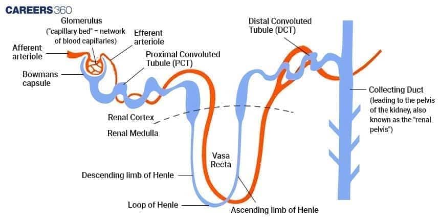
The countercurrent mechanism takes place in specific parts of the nephron. The glomerulus performs ultrafiltration, forming the initial filtrate. The loop of Henle is the main site of the countercurrent mechanism, concentrating urine by reabsorbing water and salts. The parts of a nephron are:
Glomerulus: Filtrates blood to form filtrate.
Bowman's Capsule: Collects the filtrate from the glomerulus
Proximal Convoluted Tubule: Reabsorbs nutrients, ions, and water.
Loop of Henle: Concentrates urine by the reabsorption of water and salts.
Distal Convoluted Tubule: Fine adjustment in the composition of fluid.
Collecting Duct: Finally concentrates the urine.
Countercurrent Mechanism NEET MCQs (With Answers & Explanations)
Important questions asked in NEET from this topic are:
Steps of Urine Formation
Role of Kidneys in Osmoregulation
Practice Questions for NEET
Q1. The counter-current mechanism involves ____?
Vasa recta and PCT
Vasa recta and DCT
Vasa recta and loop of Henle
Vasa recta and collecting duct
Correct answer: 3) Vasa recta and loop of Henle
Explanation:
The flow of filtrate in the two limbs of Henle’s loop is in opposite directions and thus forms a counter current. The flow of blood through the two limbs of the vasa recta is also in a counter-current pattern.
This mechanism helps to maintain a concentration gradient in the medullary interstitium, thereby concentrating the filtrate( urine).
Hence, the correct answer is option 3) Vasa recta and loop of Henle.
Q2. Among the options provided, which blood vessel in mammals contains the minimal quantity of urea?
Hepatic portal vein
Hepatic vein
Dorsal aorta
Renal vein
Correct answer: 4) Renal vein
Explanation:
The renal vein, which carries blood away from the kidneys, contains the least amount of urea among the given options. This is because the kidneys are responsible for filtering waste products, including urea from the blood. As the filtration and reabsorption processes occur in the kidneys, water and essential nutrients are reabsorbed while waste products like urea remain in the tubular fluid. Consequently, the remaining fluid, known as urine, flows out of the kidneys and into the renal vein. Compared to other blood vessels involved in filtration, such as the hepatic portal vein and hepatic vein, the renal vein has a lower concentration of urea. Thus, the renal vein contains a minimal quantity of urea.
Hence, the correct answer is option 4) Renal vein.
Q3. Mammals have the ability to produce
Isotonic urine
Hypertonic urine
Hypotonic urine
Highly alkaline urine
Correct answer: 2) Hypertonic urine
Explanation:
Mechanism of Concentration of Urine -
Mammals can produce concentrated urine.
The Henle’s loop and vasa recta play a significant role in this.
The flow of filtrate in the two limbs of Henle’s loop is in opposite directions and thus forms a counter current.
The flow of blood through the two limbs of the vasa recta is also in a countercurrent pattern.
The proximity between the Henle’s loop and vasa recta, as well as the countercurrent in them, help in maintaining an increasing osmolarity towards the inner medullary interstitium, i.e., from 300 mOsmolL–1 in the cortex to about 1200 mOsmolL–1 in the inner medulla.
This gradient is mainly caused by NaCl and urea.
NaCl is transported by the ascending limb of Henle’s loop which is exchanged with the descending limb of vasa recta.
NaCl is returned to the interstitium by the ascending portion of the vasa recta.
Similarly, small amounts of urea enter the thin segment of the ascending limb of Henle’s loop which is transported back to the interstitium by the collecting tubule.
The above-described transport of substances facilitated by the special arrangement of Henle’s loop and vasa recta is called the countercurrent mechanism.
This mechanism helps to maintain a concentration gradient in the medullary interstitium.
The presence of such an interstitial gradient helps in the easy passage of water from the collecting tubule thereby concentrating the filtrate (urine).
The overall function of the countercurrent mechanism is to concentrate the sodium chloride in the interstitial fluid and thereby cause water to diffuse out of the collecting ducts and concentrate the urine.
Human kidneys can produce urine nearly four times more concentrated than the initial filtrate formed.
Mammals produce hypertonic urine.
Hence, the correct answer is option 2) hypertonic urine.
Recommended Video on Counter Current Mechanism
Frequently Asked Questions (FAQs)
The countercurrent mechanism is a process in the nephron where fluids flow in opposite directions in the loop of Henle and vasa recta, creating and maintaining an osmotic gradient that helps concentrate urine.
It occurs mainly in the loop of Henle (counter current multiplier) and the vasa recta (counter current exchanger), with urea recycling in the collecting ducts adding to the gradient.
The descending limb allows water reabsorption, while the ascending limb actively transports NaCl, establishing the medullary osmotic gradient.
It is essential for water conservation, maintaining osmotic balance, and producing concentrated urine, which is vital for homeostasis in human physiology.
