

Micturition Reflex - Diagram and Process: Definition, Steps, Phases, Disorders
The micturition reflex is a neural feedback mechanism that coordinates the contraction of the detrusor muscle and relaxation of urinary sphincters to release urine. Stretch receptors in the bladder wall activate afferent and efferent pathways involving the spinal cord, pelvic nerves, pudendal nerve, and voluntary brain control. This guide covers storage–voiding phases, reflex steps, neural circuits, sphincter control, diagrams, FAQs, and NEET MCQs.
This Story also Contains
- What is the Micturition Reflex?
- Stages of Micturition
- Steps of the Micturition Reflex
- Role of Pudendal Nerve & Voluntary Control
- Disorders & Conditions Affecting the Micturition Reflex
- Micturition Reflex NEET MCQs (With Answers & Explanations)
- Recommended Video on Micturition
What is the Micturition Reflex?
The micturition reflex is how the body is controlled to allow urine to be released from the bladder. When the bladder is filled, the increased volume is detected by stretch receptors in the walls and will signal down the spinal cord and the brain that it requires emptying. Along with the decision to urinate, it can also be initiated by the relaxation of the internal and external sphincters of the urethra along with the contraction of the detrusor muscle of the bladder to allow urine out.
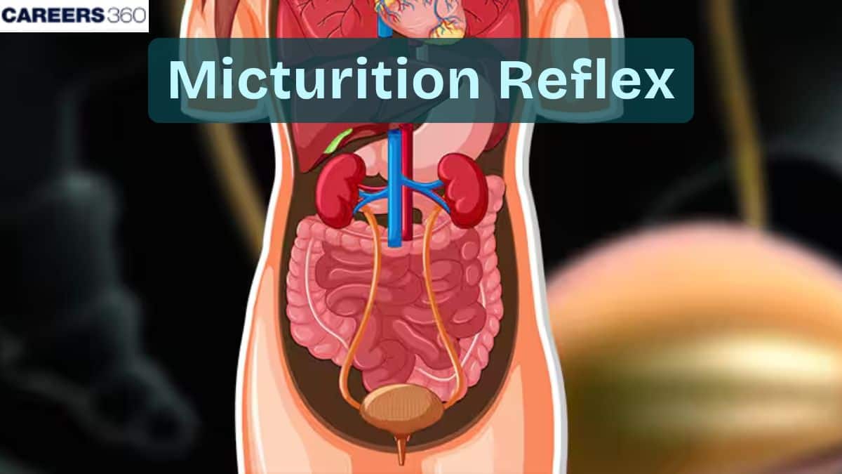
Visual Anatomy of the Reflex
It generally includes:
The bladder
The urethra
The nerves
The associated muscles
Stages of Micturition
There are two main stages of micturition:
Storage Phase
The urine is stored inside the bladder
The sphincter muscles are contracted not to let it be released.
The sympathetic nervous system stimulates the detrusor muscle to be relaxed until storage is no longer appropriate and the internal urethral sphincter stays contracted.
Voiding Phase
As the levels inside the bladder rise, the nerves get stimulated
When urination is necessary, the brain sends nerve impulses to the bladder and the urethra.
The external urethral sphincter is relaxed by the will.
Urine flows from the bladder through the urethra and out of the body.
Steps of the Micturition Reflex
Micturition is the voiding of urine from the urinary bladder.
Filling of the Urinary Bladder
It is a condition where the urinary bladder is filled with urine. When there is a filling of urine in the bladder wall of the urinary bladder is stretched.
Activation of Stretch Receptors
Stretch receptors in the urinary bladder sense the rise in its volume and form impulses which are transmitted to the nervous system. At the point of capacity of the bladder, the stretch receptors send afferent impulses through the pelvic nerve into the spinal. The stimulus in the stretch receptors produces sensory impulses which are transmitted to the central nervous system.
Afferent Impulses to Spinal Cord
The sensory impulses as a result of the stretch receptors of the bladder travel via the pelvic nerve and reach the sacral region of the spinal cord. The impulses that arrive in the bladder enter an integration centre, which is the spinal cord.
Efferent Impulses to the Bladder
Motor efferent impulses are sent from the spinal cord through the pelvic nerve back to the bladder. The efferent impulses stimulate the detrusor muscle to lead to its contraction.
Contraction of Detrusor Muscle
The contraction of the detrusor muscle increases pressure in the bladder. This pressure pushes the urine into the urethra and out of the body.
Relaxation of Internal Urethral Sphincter
There is also an opening of the internal urethral sphincter, which has an involuntary type of control. The relaxation of the involuntary sphincter will form a passage through which urine can flow from the bladder into the urethra.
Role of Pudendal Nerve & Voluntary Control
The role of pudendal nerve and voluntary control are:
Pudendal Nerve Inhibition
There shall be a stimulation of the stretch receptors within the urethra when urine flows. These stretch receptors send further afferent impulses back to the spinal cord, which also serves to reinforce the micturition reflex.
Through the relay in the spinal cord, the sensory stimulus can be transmitted further to the brain to issue commands and concomitantly inhibit signals transmitted by the pudendal nerve. Through this inhibition, it relaxes the external urethral sphincter and releases the urine.
Voluntary Control
Upon relaxation of the external sphincter, the bladder voids urine through the urethra to the external environment. This can be aided by the voluntary contractions of the musculature of the walls of the abdomen, which increase the intra-abdominal pressure that aids in the micturition.
Self-Regenerative Nature of The Reflex
The micturition reflex is self-renewable in a way that the initial contraction of the bladder causes greater stimulation of the stretch receptors. This goes on as a continuous process until the bladder has been emptied, and total micturition has been achieved.
Disorders & Conditions Affecting the Micturition Reflex
The common disorders related to micturition are:
Diseases | Symptoms | Treatment |
Urinary Incontinence |
|
|
Urinary Retention |
|
|
Neurogenic Bladder |
|
|
Micturition Reflex NEET MCQs (With Answers & Explanations)
Important questions asked in NEET from this topic are:
Mechanism of Micturition
Disorders related to Micturition
Practice Questions for NEET
Q1. Which specific action occurs in the urinary system during micturition?
Urethra relaxes
Ureter contracts
Ureter relaxes
Urethra contracts
Correct answer: 1) Urethra relaxes
Explanation:
When the bladder is full and ready to release urine, the relaxation of the urethra is crucial. It allows the urethral muscles to loosen and open up, creating a wider pathway for the urine to flow from the bladder to the outside of the body. This relaxation of the urethra is coordinated with the contraction of the bladder muscles to create a coordinated and controlled release of urine. Without the relaxation of the urethra, it would be difficult for urine to pass through and be expelled from the body.
Hence, the correct answer is option 1) Urethra relaxes.
Q2. What will happen if the stretch receptors of the urinary bladder wall are removed?
Micturition will continue
Urine will continue to collect normally in the bladder
There will be no micturition
Urine will not collect in the bladder
Correct answer: 1) Micturition will continue
Explanation:
The micturition signal is initiated by the stretching of the urinary bladder as it gets filled with urine. The stretch receptors in the bladder wall detect the increase in volume and send signals to the brain, signaling the need to urinate. The brain then sends signals to the detrusor muscle to contract, while the external sphincter muscle relaxes, allowing urine to be expelled. If the stretch receptors are removed from the bladder, the brain will not receive the signals to trigger urination, leading to a loss of control over the micturition process.
Hence, the correct answer is option 1) Micturition will continue.
Q3. The counter-current mechanism involves ____?
Vasa recta and PCT
Vasa recta and DCT
Vasa recta and loop of Henle
Vasa recta and collecting duct
Correct answer: 3) Vasa recta and loop of Henle
Explanation:
The flow of filtrate in the two limbs of Henle’s loop is in opposite directions and thus forms a counter current. The flow of blood through the two limbs of the vasa recta is also in a counter-current pattern.
This mechanism helps to maintain a concentration gradient in the medullary interstitium, thereby concentrating the filtrate( urine).
Hence, the correct answer is option 3) Vasa recta and loop of Henle.
Also Read:
Recommended Video on Micturition
Frequently Asked Questions (FAQs)
New drugs, minimally invasive surgical procedures, and sequences of ongoing clinical trials for the future appear promising.
The process by which the urinary bladder voids its urine.
The control of micturition is neural, and it is mediated by the micturition reflex, involving responses from the bladder to the brain.
It is found with some frequency that urinary incontinence, urinary retention, and overactive bladder are other common disorders and their treatments primarily include behavioural therapies, medications, and even surgery.
It generally comprises the use of urinalysis, urodynamic studies, ultrasound, and MRI.
