

Muscles: Types, Groups, Anatomy, Functions, Composition, Development
Muscles are specialized contractile tissues responsible for movement, posture, organ function, and metabolic regulation in the human body. Three major muscle types—skeletal, smooth, and cardiac—work through coordinated contraction regulated by ATP, calcium ions, and neural stimulation. This guide covers muscle types, structure, contraction mechanism, ATP–Ca²⁺ role, neuromuscular junction, disorders, diagrams, FAQs, and NEET MCQs.
This Story also Contains
- What are Muscles?
- Types of Muscles in the Human Body
- Muscle Structure
- Muscle Function: How Muscles Contract
- Types of Muscle Contraction
- Muscle Disorders and Diseases
- Muscles NEET MCQs (With Answers & Explanations)
- Recommended Video on Muscles
What are Muscles?
Muscles are specialised tissues in the human body whose function is to move by the ability to contract. They are essential for a wide range of activities within the body, such as maintaining posture, providing means and enabling locomotion, and assisting in critical functions like breathing and digestion. They contribute to overall health by way of supporting the skeletal system, protecting the internal organs, and offering help in metabolic processes.
Muscles take part in movement but play a crucial role in the overall health of the body. Regular muscle activity improves cardiovascular health, supports metabolic functions, and aids in maintaining a healthy weight. Other than daily movements, muscle strength is also vital for daily acts and the prevention of injuries of any type.
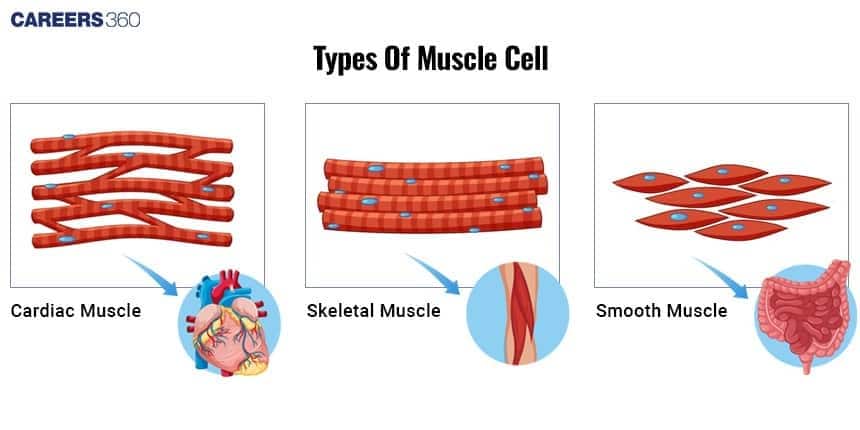
Types of Muscles in the Human Body
The types of muscles in the human body are broadly divided into three categories: skeletal, cardiac, and smooth muscles.
Skeletal Muscles
Skeletal muscles are striated in appearance.
Multinucleated fibers
Voluntary control
Fixed to bones by tendons
Extensively in limbs and torso
Cause movement due to contraction and relaxation abilities
Examples: Biceps brachii (arm), Quadriceps femoris (thigh)
Cardiac Muscles
Cardiac muscles are striated with intercalated discs
One nucleus per cell
Involuntary
In the Walls of the heart (myocardium)
Pump blood throughout the body
Contraction occurs continually in a rhythmic manner.
Smooth Muscles
Not striated
One nucleus per cell
Autonomic nervous control
In the Walls of hollow organs (e.g., intestine, blood vessels)
Move substances along the body
Examples: Muscles in the digestive tract, blood vessel walls.
Muscle Structure
Muscle structure is complex, comprising different parts that interact to produce contraction and movement.
Muscle Fibers
Elongated, cylindrical cells
Several nuclei peripherally located
Sarcomere – Functional Unit
Repeating units of myofibrils
Units responsible for muscle contraction
Myofibrils, Actin & Myosin Filaments
Myofibrils: Contractile threads within muscle fibers
Actin (thin) and myosin (thick) filaments: Proteins involved in contraction.
Connective Tissue Layers
Endomysium: surrounds individual muscle fibres
Perimysium: encases bundles of fibres (fascicles)
Epimysium: encloses the entire muscle
Tendons
Connect muscles to bones
Transmit force from muscle contraction to the skeleton.
Muscle Function: How Muscles Contract
The mechanism of muscle contraction is discussed below:
Sliding Filament Theory
Actin and myosin filaments slide past each other
Shortens the sarcomere, producing contraction.
Role of ATP
Detaches myosin head
ATP provides energy for contraction
Role of Calcium Ions (Ca²⁺)
Exposes binding sites on actin
Triggers contraction cycle
Neuromuscular Junction & Action Potential:
Synapse between a motor neuron and muscle fibre.
Action potential leads to muscle contraction.
Types of Muscle Contraction
Types of muscle contraction are:
Isotonic Contractions
Length of the muscle changes
E.g., lifting a weight
Isometric Contractions
Muscle does not change in length
E.g., holding a position like plank
Concentric Contractions
Muscle shortens when contracting
Eccentric Contractions
Muscle lengthens when contracting
Muscle Disorders and Diseases
Several disorders and diseases can compromise muscle and thus function and quality of life.
Muscular Dystrophy
Genetic mutations that alter muscle proteins.
Treatment is symptomatic and aims to retard the progression.
Duchenne muscular dystrophy: Progressive muscle weakness
Becker muscular dystrophy: Similar but milder
Myasthenia Gravis
Muscle weakness, fatigue
Autoimmune disorder at the neuromuscular junction.
Medications aimed at improving nerve-muscle communication.
Immunosuppressive therapies.
Muscle Cramps and Strains
It is caused by dehydration, overuse, and electrolyte imbalance.
This can be prevented by regular stretching and proper hydration.
Rest, ice application, compression, elevation (RICE)
Gentle stretch and rehydrate
Muscles NEET MCQs (With Answers & Explanations)
Important questions asked in NEET from this topic are:
Types of muscles
How muscles contract
Types of muscle contraction
Practice Questions for NEET
Q1. Which of the following is not a type of muscle?
Skeletal muscle
Visceral muscle
Cardiac muscle
Lymphatic muscle
Correct answer: 4) Lymphatic muscle
Explanation:
Muscles have been classified using different criteria, namely location, appearance and nature of regulation of their activities. Based on their location, three types of muscles are identified : (i) Skeletal (ii) Visceral and (iii) Cardiac. Muscles can be classified based on their location, appearance, and regulation of activity. Skeletal muscles are attached to bones and are responsible for voluntary movements. Visceral muscles, found in the walls of internal organs like the stomach and intestines, are typically involuntary and help with processes like digestion. Cardiac muscle, found in the heart, is also involuntary and specialized for rhythmic contractions. Each type of muscle has distinct structural and functional characteristics suited to its role in the body.
Hence, the correct answer is option 4) Lymphatic muscle.
Q2. Which of the following statements is incorrect ?
Smooth muscles are found in urinary bladder , alimentary canal and genital tract
A striated muscle is a syncytium i.e., a multinucleate structure
The cytoplasm of striated muscle is called endoplasm
The plasma membrane and ER of striated muscles are called sarcolemma and sarcoplasmic reticulum respectively
Correct answer: 3) The cytoplasm of striated muscle is called endoplasm
Explanation:
Visceral muscles are involuntary muscles, as their activities are not under the conscious control of the nervous system. They play a crucial role in various physiological functions, such as transporting food through the digestive tract and moving gametes through the genital tract. These muscles are primarily found in the walls of internal organs like the stomach, intestines, and blood vessels, facilitating essential bodily processes without conscious effort.
Hence the correct answer is option 3) The cytoplasm of striated muscle is called endoplasm.
Q3. The muscle fibers that contract slowly are
Red muscle fibres
White muscle fibres
Both a and b
None of these
Correct answer: 1) Red muscle fibres
Explanation:
Red muscle fibers, also known as slow-twitch fibers, contract slowly but are highly resistant to fatigue. They are rich in myoglobin, mitochondria, and blood supply, which enable sustained aerobic energy production. These fibers are well-suited for endurance activities like walking, running long distances, or maintaining posture, as they rely on oxidative metabolism for energy.
Hence, the correct answer is option 1) Red muscle fibers.
Also Read:
Recommended Video on Muscles
Frequently Asked Questions (FAQs)
Dehydration, according to some causes, muscle cramps; overuse equally causes it, and lastly, electrolyte imbalance. Prevent them by keeping hydrated, stretch your muscles often, and keep the electrolytes in balance.
The three types of muscles are skeletal, cardiac and smooth muscles.
According to the sliding filament theory, actin and myosin filaments slide past each other to shorten the muscle.
Skeletal muscles are voluntary and striated. Cardiac muscles, on the other hand, are involuntary and striated but with intercalated discs. Smooth muscles are involuntary and non-striated.
Strengthening of muscles occurs with the inclusion of resistance training, enough quantities of protein, and regular physical activities.
