

Ball and Socket Joint: Movement, Examples & Function
A ball and socket joint is a type of synovial joint in which the spherical head of one bone fits into the cup-shaped socket of another. This structural arrangement provides the widest range of movement in the human body, including rotation and movement in all planes. This guide explains the structure, movements, examples, comparison with hinge joints, clinical significance, FAQs, and NEET MCQs.
This Story also Contains
- What Is a Ball and Socket Joint?
- Structure of Ball and Socket Joint
- Functions & Movements of Ball and Socket Joint
- Diagram of Ball and Socket Joint
- Examples of Ball and Socket Joints in the Humans
- Difference Between Ball and Socket Joint and Hinge Joint
- Clinical Importance of Ball and Socket Joint
- Ball and Socket Joint NEET MCQs (With Answers & Explanations)
What Is a Ball and Socket Joint?
Ball and socket joint has a spherical end of one bone, called the ball, that fits into a rounded cavity of another bone, known as the socket. It is a type of synovial joint that enables multidirectional motion. With the high degree of mobility this arrangement provides, movements like rotation, flexion, extension, abduction, adduction and rotation are made possible.
Structure of Ball and Socket Joint
The following essential components make up the ball and socket joint:
Ball
The rounded portion of the bone that slides into the socket is called the ball. Examples in humans are the humerus (upper arm bone) and the head of the femur (thigh bone).
Socket
The structure that resembles a cup and receives the ball is called a socket. For example, the hip joint's socket is the acetabulum of the pelvis.
Articular Cartilage
The smooth substance known as articular cartilage covers the ends of bones and reduces friction when moving.
Synovial Fluid
The lubricating substance known as synovial fluid, which is present in the joint capsule, lowers friction and supports the cartilage.
Joint Capsule
A fibrous tissue that surrounds the joint and offers support and stability is called a joint capsule.
Functions & Movements of Ball and Socket Joint
The synovial fluid within the joint capsule lubricates the articular surfaces of the joints to minimise friction for smooth movement. As a result of this structure, such joints display very wide ranges of motion, enabling movements along several planes:
Flexion and Extension: Bending and straightening movements.
Abduction and Adduction: The movement away from and towards the body's midline.
Rotation: Circular movements around the central axis.
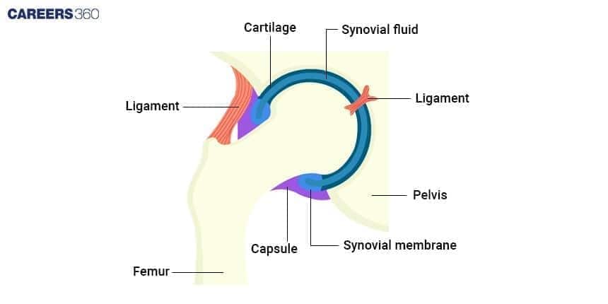
Diagram of Ball and Socket Joint
The diagram illustrates the structure of ball and socket joint in the hip joint.
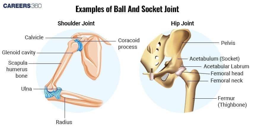
Examples of Ball and Socket Joints in the Humans
The shoulder and hip joints are the two main ball and socket joints found in the human body.
Shoulder Joint
Made up of the scapula and humerus, this joint permits a variety of motions, such as turning and raising the arms overhead.
Hip Joint
It is made up of the femur and the pelvis, and permits motions like walking, jogging, and sitting while supporting weight-bearing activities and offering stability.
Difference Between Ball and Socket Joint and Hinge Joint
Both ball and socket joint and hinge joint are synovial joints but they differ in the structure and movement. The differences between the two joints are:
Feature | Ball and Socket Joint | Hinge Joint |
Structure | Rounded head of one bone fits into a cup-like socket of another bone | Cylindrical end of one bone fits into a trough-like surface of another bone |
Movement | Multidirectional: flexion, extension, abduction, adduction, rotation | Unidirectional: mainly flexion and extension |
Mobility | Highest mobility | Limited mobility |
Examples in humans | Shoulder joint, Hip joint | Elbow joint, Knee joint, Interphalangeal joints |
Clinical Importance of Ball and Socket Joint
The clinical importance of ball and socket joint is:
Shoulder Dislocation
The shoulder joint, though highly mobile, is prone to dislocation due to a shallow socket (glenoid cavity).
Hip Joint Fractures
Common in elderly people with osteoporosis, especially fracture of the femoral neck.
Arthritis
Both shoulder and hip joints are affected by osteoarthritis and rheumatoid arthritis, causing pain and stiffness.
Sports Injuries
Frequent in athletes due to overuse or trauma, affecting shoulder stability and hip strength.
Ball and Socket Joint NEET MCQs (With Answers & Explanations)
Important questions asked in NEET from this topic are:
Structure of ball and socket joint
Examples of ball and socket joint in human
Practice Questions for NEET
Q1. Which of the following is/are not correctly matched pairs?
(i) Ball and socket joint - Between humerus and pectoral girdle
(ii) Pivot joint - Between carpal and metacarpal
(iii) Saddle joint - Between atlas and axis
(iv) Gliding joint - Between the carpals
(v) Fibrous joint - In flat skull bones
(ii) and (iii)
(i) and (iv)
(v) only
(ii) only
Correct answer: (ii) and (iii)
Explanation:
(i) Ball and socket joint - Between humerus and pectoral girdle: This is correct. The shoulder joint is a ball and socket joint where the head of the humerus fits into the socket of the scapula (part of the pectoral girdle).
(ii) Pivot joint - Between carpal and metacarpal: This is incorrect. The joint between the carpal and metacarpal bones is a saddle joint, not a pivot joint. A pivot joint is found between the atlas and axis vertebrae in the neck (which allows for rotation of the head).
(iii) Saddle joint - Between atlas and axis: This is incorrect. The joint between the atlas and axis vertebrae is a pivot joint, not a saddle joint. A saddle joint is found in areas like the thumb (between the carpal and metacarpal bones).
(iv) Gliding joint - Between the carpals: This is correct. The joints between the carpals (wrist bones) are gliding joints allowing for limited sliding movements.
(v) Fibrous joint - In flat skull bones: This is correct. The bones in the skull are joined by fibrous joints called sutures which are immovable.
Hence, the correct answer is option 1) (ii) and (iii).
Q2. The pivot joint between the atlas and the axis is a type of:
Fibrous Joint
Cartilaginous Joint
Synovial Joint
Saddle Joint
Correct answer: 3) Synovial Joint
Explanation:
The pivot joint connecting the atlas (C1) and axis (C2) vertebrae is a unique synovial joint, commonly referred to as the atlantoaxial pivot joint. It is essential for the head's rotational movement, such as when one nods in disagreement or affirmation. This joint's structure includes the odontoid process (dens) of the axis, which serves as the pivot, while the anterior arch of the atlas and the transverse ligament provide stability and keep the dens in position. The primary function of this joint is to allow the atlas to rotate around the dens, facilitating the head's rotation. It is a key component of the cervical spine, contributing significantly to neck flexibility and overall body coordination. It is crucial to recognize that pivot joints are designed for rotation around a single axis, and the atlantoaxial joint exemplifies this, playing a pivotal role in head and neck mobility.
Hence, the correct answer is option 3) Synovial joint.
Q3. Match the following columns
Column I (Types of Synovial) | Column II (Bones Involved) | ||
A | Ball and Socket | 1 | Carpal and metacarpal of thumb |
B | Hinge | 2 | Atlas and axis |
C | Pivot | 3 | Humerus and pectoral girdle |
D | Saddle | 4 | Knee |
A-3, B-4,C-2, D-1
A-1, B-4, C-3, D-2
A-2, B-3, C-4, D-1
A-3, B-4, C-1, D-2
Correct answer: 1) A-3, B-4,C-2, D-1
Explanation:
Hinge Joint: The convex surface of one bone articulates with the concave surface of another. Up-and-down motion in one plane is possible. Examples: the elbow and knee joints.
Ball-and-socket joint: The ball-shaped head of one bone fits into the cup-shaped socket of another. Movement in all planes, as well as rotation, is possible. Examples: the shoulder and hip joints.
Saddle joint: Each bone is saddle-shaped and fits into the complementary regions of the other. A variety of movements are possible. Example: the joint between the carpal and metacarpal bones of the thumb.
Pivot joint: A small, cylindrical projection of one bone pivots within the ring formed of bone and ligament of another bone. Only rotation is possible. Examples: the joint between the proximal ends of the radius and ulna, and the joint between the atlas and axis.
Hence, the correct answer is option 1) A-3, B-4,C-2, D-1.
Also Read:
Frequently Asked Questions (FAQs)
The ligament stabilises the joints and tendons attach muscles to bones, enabling movement and muscle contraction.
Ball and socket joints allow for multidirectional movements in all directions and rotation. Hinge joints essentially have flexion and extension along a single axis.
Among others—joint dislocations, rotator cuff tears, and hip fractures—are typical injuries. These often require medical intervention and subsequent rehabilitation.
It is observed that with ageing there is a degeneration of joints thus increasing the chances of disorders such as osteoarthritis, which severely affects mobility and causes pain.
