

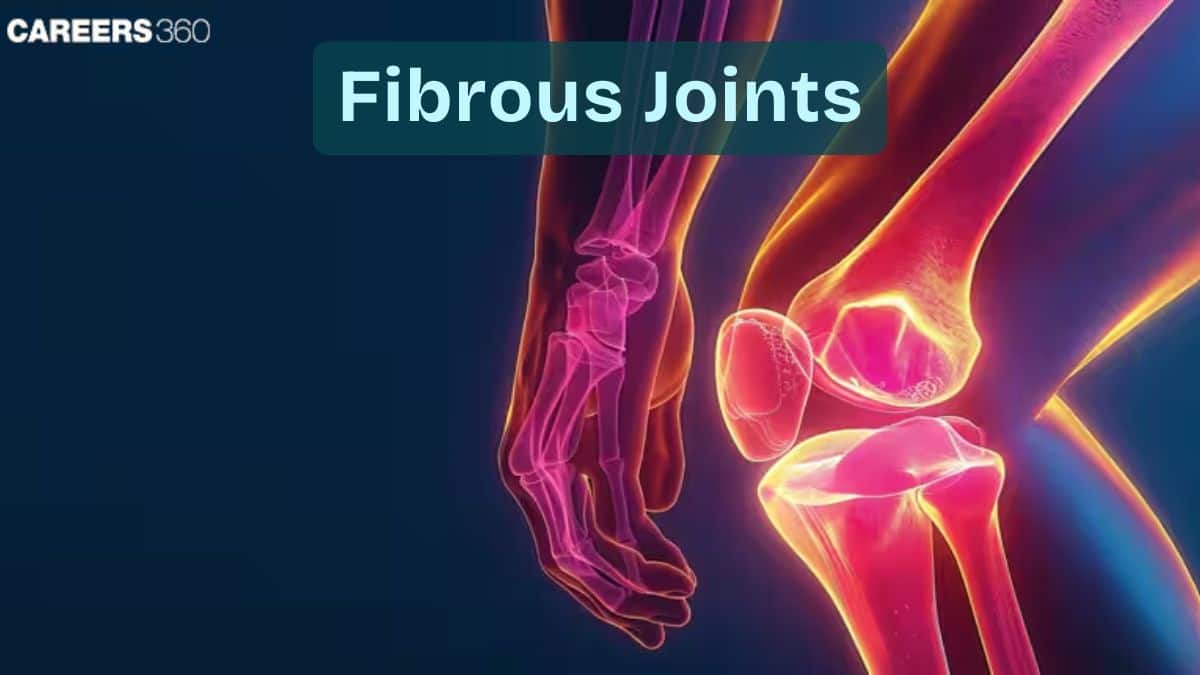
Fibrous Joints: Definition, Meaning, Examples, Types, Diagram, Classification
Fibrous joints are immovable or slightly movable joints in which bones are connected by dense fibrous connective tissue rich in collagen fibres. These joints provide stability and structural integrity to the skeletal system rather than mobility. This guide explains the definition, types, examples, structure, clinical importance, FAQs, and NEET-focused MCQs on fibrous joints.
This Story also Contains
- What are Fibrous Joints?
- Characteristics of Fibrous Joints
- Types of Fibrous Joints
- Clinical Significance of Fibrous Joints
- Fibrous vs Cartilaginous vs Synovial Joints
- Fibrous Joints NEET MCQs (With Answers & Explanations)
What are Fibrous Joints?
Fibrous joints, also known as immovable joints, are directly connected by dense fibrous connective tissue, mostly made of collagen fibres. Synovial joints allow for great movement, but fibrous joints are designed to offer stability and very little or no movement. These do not have a cavity and are important in maintaining the structural integrity of the body.
Examples of fibrous joints are:
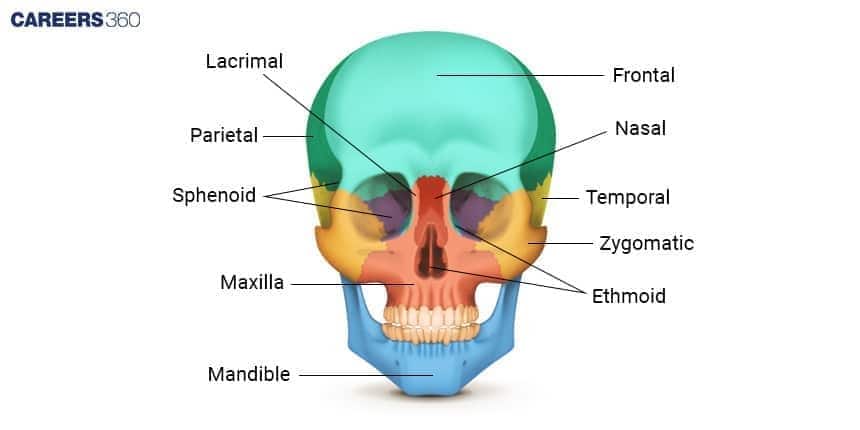
Suture Joint: These are found in the skull, where bones interlock closely and form an immovable connection. This is another example of an immovable joint.
Gomphosis Joint: That joint between the teeth and their sockets in the jawbone secured by the periodontal ligament.
Characteristics of Fibrous Joints
The characteristics of fibrous joints are joints in which bones are held together by dense fibrous connective tissue, providing strength and stability rather than movement.
The bones are tightly joined by fibrous connective tissue, ensuring firm attachment.
These joints allow minimal or no movement, making them functionally immovable.
Their rigid structure provides great mechanical strength and resistance to stress.
Fibrous joints like skull sutures protect organs like the brain by forming a strong protective casing.
Types of Fibrous Joints
The different types of fibrous joints are:
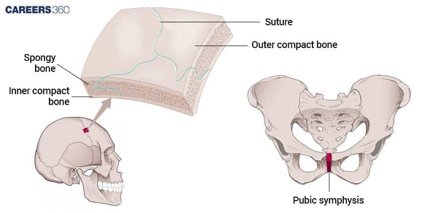
Sutures
Sutures exist only in the skull, where the bones are anatomically tightly set together with a thin layer of fibrous tissue. An immovable joint is formed, which helps protect the brain and maintain the shape of the skull. Examples of such sutures are the sagittal suture and lambdoid suture.
Gomphosis
Gomphosis refers to specialized fibrous joints that connect the teeth to their sockets in the maxilla and mandible. The fibrous connection here is quite strong in preventing loose attachment, hence the firmness of teeth for every action of chewing or doing anything with the oral cavity.
Syndesmosis
In syndesmosis joints, the bones are more widely spaced and are ligamentous or by interosseous membrane held in place. Examples include the distal ends of the forearm radius and ulna and of the lower leg tibia and fibula. These joints will allow some movement, though little, like rotation at the elbow or shock absorptive movement at the knee from the impact of walking.
Clinical Significance of Fibrous Joints
Fibrous joints, also called immovable or fixed, are crucial in orthopaedics and dentistry, and injuries, such as dislocation or fractures through syndesmosis should be treated very carefully with the aim of restoration of function to prevent further long-lasting problems.
They exist to stabilize and support most parts of the body in either a simple or a comprehensive framework. Thus, the physician can only do so much while operating within normal physiology. Any explanation of these structures will rely on diagrams of the several types of fibrous joints, such as suture and gomphosis as well as fixed joint diagram types.
Fibrous vs Cartilaginous vs Synovial Joints
Based on the movement the joints are divided into fibrous, cartilaginous and synovial joints. The difference between these is discussed in the table below:
Feature | Fibrous Joints | Cartilaginous Joints | Synovial Joints |
Connecting tissue | Dense fibrous tissue | Synovial cavity with synovial fluid | |
Movement | No movement | Slight movement | Free movement |
Strength | High | Moderate | Less than fibrous |
Function | Stability and protection | Support with limited flexibility | Wide range of movements |
Examples | Skull suture | Pubic symphysis | Knee, shoulder |
Fibrous Joints NEET MCQs (With Answers & Explanations)
Important questions asked in NEET from this topic are:
Classification of fibrous joints
Fibrous vs Cartilaginous vs Synovial joints
Practice Questions for NEET
Q1. Which one of the following pairs is correctly matched?
Cartilaginous joint - Skull bones
Hinge joint - Between vertebrae
Fibrous joint - between phalanges
Gliding joint - between the bones of the wrist
Correct answer: 4) Gliding joint - between the bones of the wrist
Explanation:
Cartilaginous joints are those in which cartilage connects the bones, as in the ribs to the sternum or the spine between vertebrae. Therefore, it does not match the skull bones correctly.
Hinge joints: These joints, which are located in areas like the elbow and knee rather than in between vertebrae, provide movement in a single plane, much like a door hinge.
Fibrous joint: These joints are made up of bones that are joined by fibrous tissue, not between phalanges, as in the case of the skull's sutures.
Gliding joint: These joints, which are located in between the wrist's bones (such as the carpals), permit sliding or gliding motions. This is the appropriately matched pair as a result.
Hence, the correct answer is option 4) Gliding joint - between the bones of the wrist
Q2. The pivot joint between the atlas and the axis is a type of:
Fibrous joint
Cartilaginous joint
Synovial joint
Saddle joint
Correct answer: 3) Synovial joint
Explanation:
The pivot joint connecting the atlas (C1) and axis (C2) vertebrae is a unique synovial joint, commonly referred to as the atlantoaxial pivot joint. It is essential for the head's rotational movement, such as when one nods in disagreement or affirmation. This joint's structure includes the odontoid process (dens) of the axis, which serves as the pivot, while the anterior arch of the atlas and the transverse ligament provide stability and keep the dens in position. The primary function of this joint is to allow the atlas to rotate around the dens, facilitating the head's rotation. It is a key component of the cervical spine, contributing significantly to neck flexibility and overall body coordination. It is crucial to recognize that pivot joints are designed for rotation around a single axis, and the atlantoaxial joint exemplifies this, playing a pivotal role in head and neck mobility.
Hence, the correct answer is option 3) Synovial joint.
Q3. The elbow joint is an example of:
Hinge joint
Gliding joint
Ball and socket joint
Pivot joint
Correct answer: 1) Hinge joint
Explanation:
One type of synovial joint, also referred to as a freely moving joint, is the hinge joint. The elbow and knee both have hinge joints. The pivot joint is located between the atlas and axis, the gliding joint is located between the carpals and the ball and socket joint is located between the shoulder and hips.
Hence, the correct answer is option 1) Hinge joint.
Also Read:
Frequently Asked Questions (FAQs)
Amphiarthrosis describes those joints that allow only slightly more motion between the bones. Examples include syndesmoses, which are interosseous membrane equations between the radius and ulna.
No, sutures are not found in the mandible joints because the mandible is the only movable bone in the skull used during operations such as talking and eating.
Synarthrosis refers to an immovable joint where simple motion is impossible. Examples are skull sutures, anchorage of teeth with the cavity of the bone, and syndesmoses between the tibia and fibula.
