

Kidney and Nephron – Diagram, Definition, Function, Structure, Facts
Kidneys are vital excretory organs that filter blood, eliminate wastes, regulate fluid balance, and maintain homeostasis through nearly one million nephrons per kidney. Each nephron performs filtration, reabsorption, and secretion through its structural components—glomerulus, Bowman’s capsule, PCT, Loop of Henle, DCT, and collecting duct. This guide covers kidney structure, nephron types, functions, diagrams, urine concentration mechanisms, related disorders, FAQs, and NEET MCQs.
This Story also Contains
- What Is the Kidney? (Definition & Importance)
- Structure of Human Kidney
- Nephron – Functional Unit of Kidney
- Types of Nephrons
- Mechanism of Urine Formation (Three Steps)
- Disorders of the Excretory System
- Kidney & Nephron NEET MCQs (With Answers & Explanations)
- Recommended Video for Kidney and Nephron
What Is the Kidney? (Definition & Importance)
The kidneys do the major work of the urinary system. The other parts of the system are mainly passageways and storage areas. The paired kidneys are reddish, kidney bean–shaped organs located between the peritoneum and the posterior wall of the abdomen. The right kidney is slightly lower than the left because the liver occupies considerable space on the right side superior to the kidney.
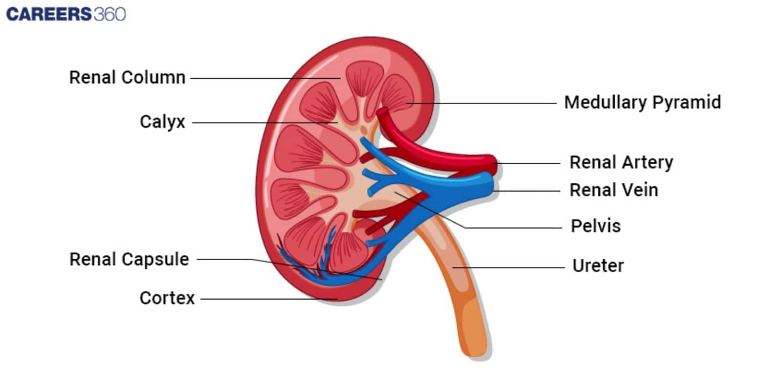
Structure of Human Kidney
The anatomy of the kidney is discussed below:
Shape and Size
The kidneys are paired, bean-shaped organs, each being about 11-14 cm long, 6 cm wide and 4 cm in thickness. They weigh about 150 grams in adults.
Position
One on either side of the spine in the abdominal cavity, the kidneys lie directly below the rib cage. A little lower than the left, the right kidney is normally located to provide space for the bulky liver.
Renal Cortex
The outer part of the kidney, having renal corpuscles and the bulk of renal tubules. It is said to be the real machine for the filtration of blood and the place of urine formation.
Renal Medulla
It is the inner portion that contains the renal pyramids. The renal columns are the visible portions of tissue between the pyramids. The renal pyramids are made of the Henle loops and collecting ducts, elements that are responsible for concentrating the urine and transporting it.
Renal Pelvis
The hollow spelt in the centre of each kidney collects urine from medullary pyramids and leads it into the ureter. So, it works as a funnel passage of urine to the bladder.
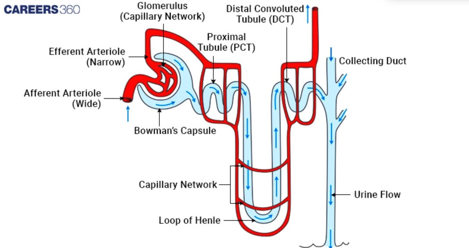
Nephron – Functional Unit of Kidney
Nephrons are the functional units of the kidneys. Each nephron consists of two parts: a renal corpuscle, where blood plasma is filtered, and a renal tubule into which the filtered fluid passes.
Renal Corpuscle
Glomerulus: Ball of capillaries where filtration of blood starts. The pressure there of the capillaries pushes water, ions and small molecules out into Bowman's capsule.
Bowman's Capsule: A cup-shaped chamber surrounding that glomerulus which gathers the filtrate from the glomerulus.
Renal Tubule
Proximal Convoluted Tubule (PCT): This is the first section of the renal tubule of the nephron. Here, approximately 65% of filtered water and 65% of the filtered ions are reabsorbed.
Loop of Henle: This U-shaped loop projects into the medulla. Water and salt are reabsorbed there, concentrating the urine. It is composed of two limbs. There is a downrunning limb, which is permeable to water, and an uprunning limb, which is permeable to salts.
Distal Convoluted Tubule (DCT): This is followed by additional fine-tuning of the filtrate due to differential reabsorptions and secretion—chiefly under aldosterone influence.
Collecting Duct: It is formed by a collection of many nephrons; this at last completes the concentration of the urine and hence carries it out to the renal pelvis.
Types of Nephrons
The basic functional unit of the kidney is the nephron, which filters the blood of waste. The different types of nephron are:
Cortical Nephron
This type of nephron mostly lies in the renal cortex. They have been found with their shorter loops of Henle which lead to most of the renal bulk filtration and absorption.
Juxtamedullary Nephron
They too have long loops that dip deep into the medulla from a position close to the junction of the cortex and medulla.
Mechanism of Urine Formation (Three Steps)
The net process of urine formation comprises filtration, reabsorption, and secretion. This occurs in the kidneys.
Glomerular Filtration
Blood enters the glomerulus water and solutes get filtered into Bowman's capsule.
Filtration is driven by blood pressure.
Tubular Reabsorption
Essential substances like glucose, amino acids, and ions get reabsorbed into the blood.
Basically in the proximal convoluted tubule.
Plays a significant role in the reabsorption process of important nutrients and water balance.
Tubular Secretion
The secretion of waste products hydrogen ions, potassium, and some drugs into the tubule.
This contributes to excess unwanted compounds that are eliminated from the blood.
Disorders of the Excretory System
Many disorders of the excretory system, which inhibits the proper clearance of wastes and continued homeostasis.
Kidney Stones
These are caused by low fluid intake, high salt consumption, and genetics.
Symptoms include severe pain, blood in urine, and frequent urination.
Treatment is increased fluid intake, medications, and surgery.
Renal Failure
The kidneys lose their ability to filter blood effectively, causing waste products and toxins to accumulate.
Fluid, electrolyte, and acid–base balance become disturbed, affecting multiple organ systems.
Severe cases may require dialysis or kidney transplantation to sustain life.
GFR Abnormalities
Stages 1 to 5, based on GFR
Caused by diabetes, blood pressure, glomerulonephritis.
Symptoms include fatigue, swelling, nausea, and decreased urine output.
Treatment options include behavioural changes, medications, dialysis, and kidney transplants.
Kidney & Nephron NEET MCQs (With Answers & Explanations)
Important questions asked in NEET from this topic are:
Major organs in excretory system
Mechanism of urine formation
Practice Questions for NEET
Q1. Middle protective layer seen in kidneys of human is
Renal capsule
Renal fascia
Renal peritoneum
Adipose layer
Correct answer: 4) Adipose layer
Explanation:
The layers surrounding the kidney are three.
a) Renal fascia (outermost) - This is the dense connective tissue layer that provides support and protection to the kidneys and adrenal glands.
b) Adipose capsule (intermediate) - This is known as perirenal fat; it surrounds the kidneys and serves to cushion and insulate them.
c) Renal capsule (innermost) - A thick fibrous layer that directly encases the kidney, providing further protection and maintaining its shape.
Hence, the correct answer is option 4) Adipose layer
Q2. Columns of Bertini in the kidneys of mammals are formed as extensions of
Cortex in the medulla
Cortex in pelvis
Medulla in pelvis
Pelvis in ureter
Correct answer: 1) Cortex in the medulla
Explanation:
Columns of Bertini-The renal cortex extends between the renal pyramids in the kidney to form the columns of Bertini, also referred to as renal columns. They consist of fibrous material, urine tubes, and blood arteries. The cortex is anchored in place by the Bertini columns.
Hence, the correct answer is option 1) Cortex in the medulla.
Q3. Skin can eliminate certain substances like
Sweat
NaCl
Urea
All of these
Correct answer: 4) All of these
Explanation:
The sweat and sebaceous glands in the skin can eliminate certain substances through their secretions. The sweat produced by the sweat glands is a watery fluid containing NaCl, small amounts of urea, lactic acid, etc. This procedure aids in both thermoregulation and the body's removal of trace amounts of waste.
Hence, the correct answer is option 4)All of these.
Also Read:
Recommended Video for Kidney and Nephron
Frequently Asked Questions (FAQs)
The major processes through which the nephron starts producing urine are:
Glomerular Filtration
Tubular Reabsorption
Tubular Secretion
Common kidney disorders include:
Chronic Renal Failure
Acute Kidney Injury
Kidney Stones
Urinary Tract Infections
Polycystic Kidney Disease
For Healthy Kidneys:
Remain Hydrated
Healthy Diet
Regular Exercise
Avoid smoking and excessive alcohol consumption
The kidney's primary role is to filter the blood to remove waste products, excess fluid and electrolytes, which are passed in urine and thus excreted out of the body.
A nephron consists of a renal corpuscle and the renal tubule which forms the functional unit of the kidney.
