

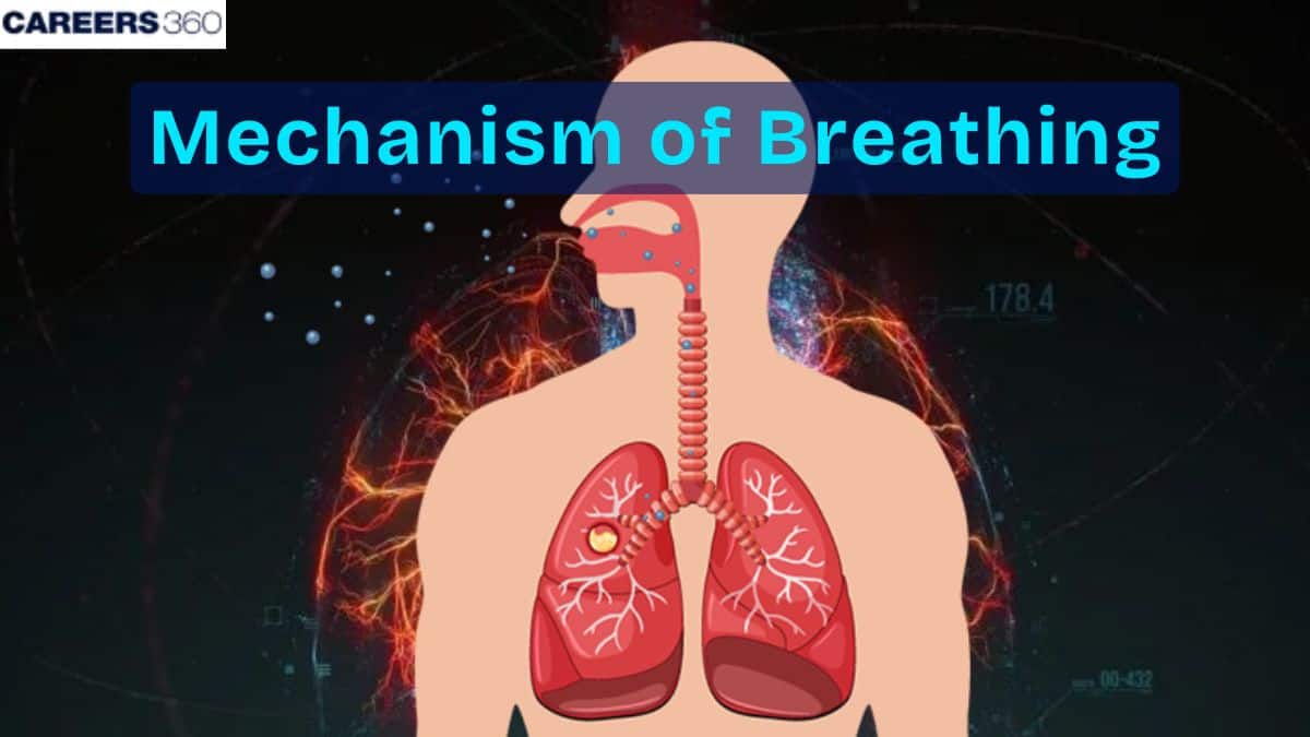
Mechanism of Breathing: Definition, Diagram, Functions
The mechanism of breathing includes the coordinated actions of the diaphragm, intercostal muscles, and thoracic cavity changes during inhalation and exhalation. Inspiration involves expansion of the thoracic cavity creating negative pressure, while expiration occurs when the cavity relaxes and pressure increases. Understanding these steps is essential for NEET, Class 11 Biology, and human physiology.
This Story also Contains
- What Is the Mechanism of Breathing?
- Inhalation (Inspiration) – Step-by-Step Process
- Exhalation (Expiration) – Step-by-Step Process
- Muscles Involved in Breathing
- Lung Compliance and Elasticity
- Airway Resistance
- Intrapleural Pressure and Negative Pressure Breathing
- Gas Transport During Respiration
- Difference Between Inhalation and Exhalation
- Mechanism of Breathing NEET MCQs (With Answers & Explanations)
- Recommended Video for "Mechanism of Breathing"
What Is the Mechanism of Breathing?
Respiration is the physiological exercise through which gases, mainly oxygen and carbon dioxide are exhaled through the lungs of an organism. This is important for cellular respiration as this moves the oxygen required in aerobic respiration to reach the cells and at the same time takes out carbon dioxide, a waste product. Proper functioning of the lungs guarantees a constant supply of oxygen to the cells for their metabolism and regulates the proper distribution of gases, which are significant for the life process.
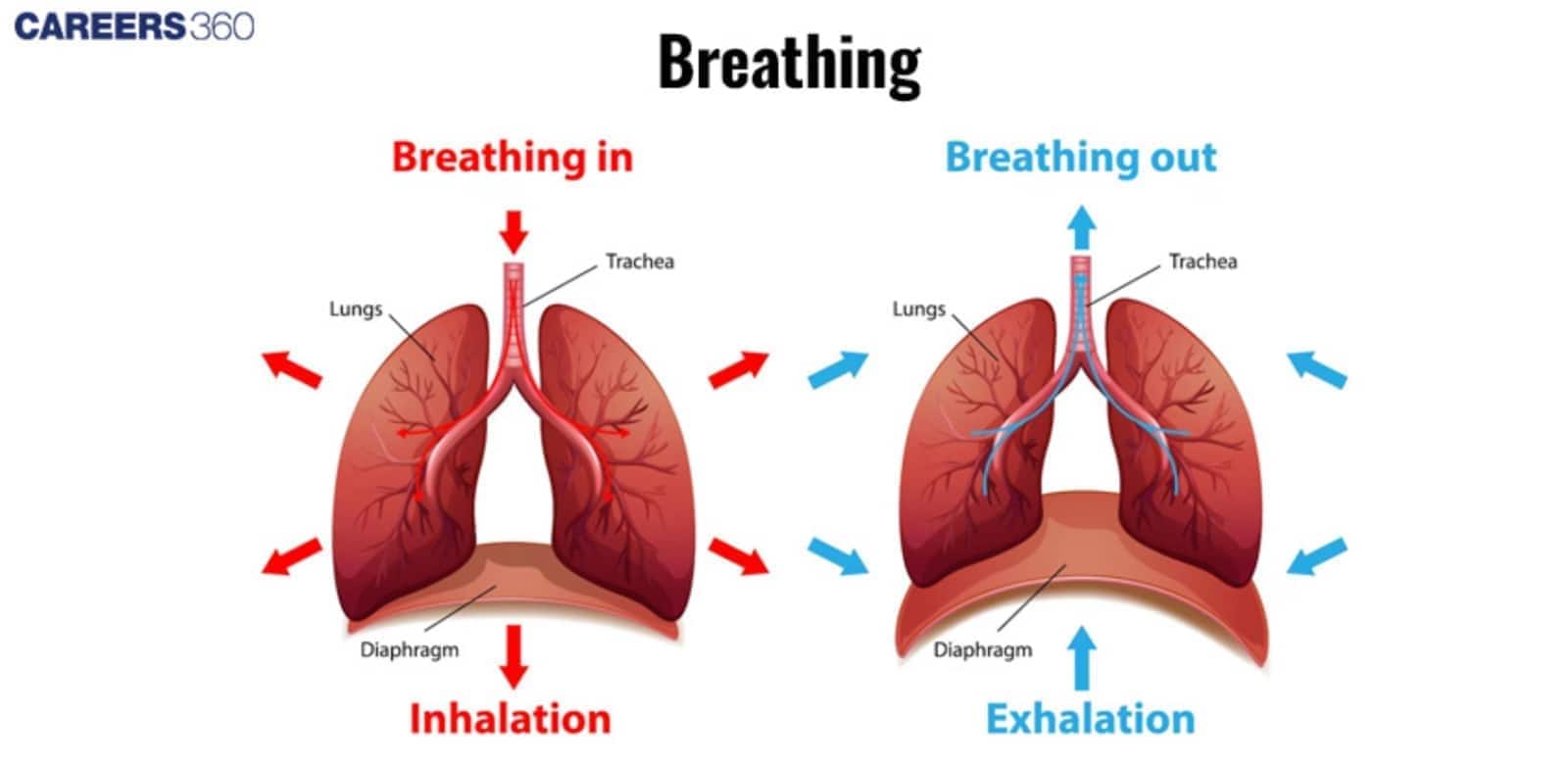
Inhalation (Inspiration) – Step-by-Step Process
Inhalation is characterised by the contraction of the diaphragm and the intercostal muscles to raise the ribs and expand the thoracic cavity. This contraction generates a vacuum, or negative pressure within the thoracic cavity and air rushes into the lungs.
During the process of inhalation, contraction of the muscles surrounding the thoracic cavity increases the volume of this cavity and thus forces the air through the respiratory tract and into the alveoli to participate in the exchange of gases.
Exhalation (Expiration) – Step-by-Step Process
Exhalation takes place when the diaphragm and intercostals decrease their size, decreasing the volume of the thoracic cavity. This relaxation leads to positive pressure in the thoracic cavity which in turn forces air out of the lungs and out of the respiratory tract. The reduction of volume ejects the air laden with carbon dioxide ending the breathing cycle.
Muscles Involved in Breathing
The muscles involved in breathing:
Diaphragm
The diaphragm functions as a muscle which is in the shape of a dome and is situated at the bottom part of the thoracic cavity.
Inhalation is done by the contraction of the diaphragm, which becomes flattened. By doing so, the pressure in the thoracic cavity is reduced enabling air to enter the lungs.
Intercostal Muscles
The intercostal muscles are located in between the ribs of an individual’s body.
These muscles contract during inhalation and, at the same time, they relax during exhalation to narrow the thoracic cavity hence forcing air out of the lungs.
Lung Compliance and Elasticity
Lung compliance deals with the ease with which the lungs can expand and dilate given some pressure.
High lung compliance means that the lungs can be expanded easily and can therefore fill up easily while low compliance means that the lungs are stiff and cannot be filled easily.
Accessibility of the lungs can be affected by the degree of elasticity of the lung tissues, and the quantity and nature of the surface tension-reducing agents. This leads to disorders in respiration.
Lung compliance is an index of work that the respiratory muscles need to do to achieve a certain level of ventilation.
Airway Resistance
Airway resistance is the opposition force within the respiratory tracts to the flow through them.
This depends on the dimensions like the diameter of the airway, mucus production, and bronchial constriction.
There is always the case of high resistance, this is because of the constriction of the tubes through which air has to pass, or the presence of an obstructing factor that makes it difficult to breathe.
It is important to deal with airway resistance if breathing properly and if fresh and expired air is going to get to the lungs.
Intrapleural Pressure and Negative Pressure Breathing
The pressure in the pleural cavity, the space between the lungs and pleura, is referred to as intrapleural breathing. This pressure is less than atmospheric pressure, known as negative pressure, which is vital in the mechanics of respiration.
The transpulmonary pressure or the pressure difference is accountable for the lung movement during breathing. The intrapleural pressure becomes more negative during inhalation and causes the lungs to expand. During expiration, the pressure increases, causing the lungs to recoil.
The lungs' elasticity and the surface tension of alveolar fluid pull the lungs inward, while the thoracic wall and pleural fluid create an opposing force. This balance results in negative intrapleural pressure, a key concept in the mechanism of breathing.
Gas Transport During Respiration
Respiratory gas transport refers to the transport of oxygen and carbon dioxide by the blood. Oxygen-rich blood from the lungs flows to the heart through pulmonary veins. The heart pumps this oxygenated blood to the rest of the body through the aorta. At the same time, deoxygenated blood with carbon dioxide is returned to the lungs by the pulmonary arteries for gas exchange. The whole process is repeated continuously so that oxygen, carbon dioxide, and carbon dioxide balance are maintained in the body. The gas exchange in the lungs involves the following:
Diffusion in Alveoli
In the alveoli oxygen from the inhaled air moves passively through the walls of the alveoli into the blood in capillaries, and at the same time, carbon dioxide diffuses active from the blood into the alveoli and thus is exhaled.
This exchange is driven by partial pressure gradients. Oxygen diffuses from the place that has high partial pressure in the alveoli relative to that found in blood and similarly for the diffusion of carbon dioxide.
Hemoglobin and Oxygen Transport
Oxygen on reaching the lungs gets attached to the haemoglobin molecules and forms oxyhemoglobin which is easily transported to the tissues.
The oxygen dissociation curve shows how the affinity of haemoglobin for oxygen varies with different concentrations of oxygen partial pressure.
At high partial pressures, for example in the lungs, haemoglobin grabs more oxygen than from the lower pressure found in tissues where oxygen is needed, for use by the cells.
Difference Between Inhalation and Exhalation
The difference between inhalation and exhalation is included in the table below:
Feature | Inhalation | Exhalation |
Muscle action | Diaphragm contracts | Diaphragm relaxes |
Rib movement | Ribs move up and out | Ribs move down and in |
Thoracic volume | Increases | Decreases |
Pressure | Decreases (negative) | Increases (positive) |
Air movement | Into the lungs | Out of the lungs |
Mechanism of Breathing NEET MCQs (With Answers & Explanations)
Important topics for NEET are:
Inhalation and exhalation
Muscles involved in breathing
Intrapleural pressure
Practice Questions for NEET
Q1. Breathing involves:
Inspiration
Respiration
Expiration
Both 1 and 3
Correct answer: 4) Both 1 and 3
Explanation:
Breathing is the process of air exchange between the atmosphere and the lungs, consisting of two main stages:
Inspiration: During inspiration, the diaphragm contracts and moves downward, while the intercostal muscles lift the ribcage, increasing the thoracic cavity's volume. This reduces the pressure inside the lungs, causing atmospheric air to flow into the alveoli.
Expiration: During expiration, the diaphragm relaxes and moves upward, and the intercostal muscles lower the ribcage, decreasing the thoracic cavity's volume. This increases the pressure inside the lungs, forcing the alveolar air out into the atmosphere.
These processes are vital for oxygen intake and carbon dioxide removal, ensuring efficient gas exchange in the body.
Hence, the correct answer is option 4) Both 1 and 3.
Q2. What is the significance of the negative pressure in human lungs during inspiration?
It helps to push air into the lungs
It creates a pressure gradient for the movement of air into the lungs
It prevents the collapse of the lungs
It has no significant role in the process of breathing
Correct answer: 2) It creates a pressure gradient for the movement of air into the lungs
Explanation:
The contraction of the diaphragm and intercostal muscles in the human expands the thoracic cavity, producing negative pressure inside the lungs during inspiration. This makes the alveolar pressure lower than the atmospheric pressure. This causes the pressure gradient between the atmosphere and the alveoli, which draws air into the lungs. When the air enters the lungs, the pressure inside and outside the lungs becomes equal. This facilitates the efficient exchange of gases when breathing.
Hence, the correct answer is option 2) It creates a pressure gradient for the movement of air into the lungs.
Q3. Mark the correct pair of muscles involved in the normal breathing in humans
External and internal intercostal muscles
Diaphragm and abdominal muscles
Diaphragm and external intercostal muscles
Diaphragm and intercostal muscles
Correct answer: 4) Diaphragm and intercostal muscles
Explanation:
The diaphragm and intercostal muscles are essential for normal breathing in humans, facilitating the movement of air into and out of the lungs. During inhalation, the diaphragm contracts and flattens, increasing thoracic cavity volume, while the external intercostal muscles pull the ribs upward and outward, creating a pressure gradient that allows air to enter. During exhalation, the diaphragm relaxes and returns to its dome shape, and the intercostal muscles relax, reducing thoracic cavity volume and expelling air. This coordinated action ensures efficient oxygen intake and carbon dioxide removal.
Hence, the correct answer is option 4) Diaphragm and intercostal muscles.
Also Read:
Recommended Video for "Mechanism of Breathing"
Frequently Asked Questions (FAQs)
The mechanism of breathing is the process by which air moves in and out of the lungs. It involves two main phases: inhalation (breathing in) and exhalation (breathing out).
The diaphragm is a conical muscle which splits the thoracic cavity from the abdominal cavity. When the person breathes in, it relaxes and becomes thin to expand the volume of the thoracic cavity and produce a negative pressure that sucks in air into the lungs.
Inspiration is the process of taking in air into the lungs. Expiration is the process of the removal of air from the lungs.
This is through the respiratory centre in the brainstem, especially the medulla oblongata and pons. These centres check the levels of the concentration of carbon dioxide, amount of oxygen and blood pH and modify the depth and frequency of breath.
Some of the diseases affecting the respiratory system are asthma, COPD, chronic bronchitis, pneumonia and pulmonary fibrosis.
