

Structure of Ear: Facts, Function, Parts
The human ear performs two essential functions—hearing and balance—through its outer, middle, and inner structural divisions. Sound waves are collected, amplified, and converted into electrical impulses by the cochlea, while the vestibular apparatus maintains equilibrium. This guide explains the anatomy, physiology, pathways, equilibrium mechanism, diagrams, and NEET-focused MCQs.
This Story also Contains
- Introduction — Ear as Hearing + Balance Organ
- Outer Ear (External Ear)
- Middle Ear
- Inner Ear (Labyrinth)
- Cochlea — Organ of Hearing
- Vestibular Apparatus (Equilibrium Organ)
- Hearing Mechanism
- Mechanism of Equilibrium
- Human Ear Diagram NEET MCQs (With Answers & Explanations)
- Recommended Video on the Structure of Ear
Introduction — Ear as Hearing + Balance Organ
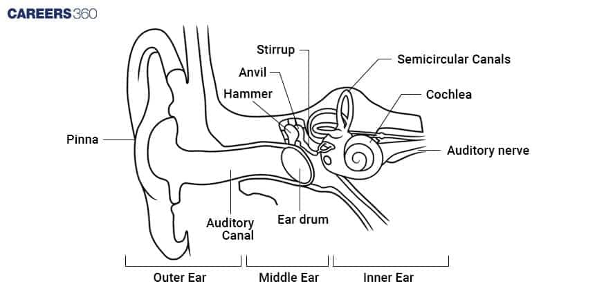
The human ear consists of two functions, hearing and balance. Three elements make up the human ear, the outer ear collects the sound, the middle ear amplifies it, and the inner ear translates it into electrical signals that are sent to the brain. The inner ear also contains structures associated with balance. Put simply, the human ear helps us hear and balance ourselves.
Outer Ear (External Ear)
Anatomy of the ear's outer or external parts includes the following:
Pinna
Small hairs and glands on the outermost part secrete sticky wax and will avoid the entry of foreign organisms and dust into the ear. It will receive the sound.
External Auditory Canal (Meatus)
Runs from the pinna to the tympanic membrane, packed with wax glands.
Tympanic Membrane
Made of connective tissue, it is covered on the outside by skin and is lined with a mucous membrane on the inside. This membrane separates the external ear from the middle ear.
Middle Ear
The middle ear contains the following anatomy:
Ossicles
A chain of three tiny bones: malleus, incus, and stapes.
Malleus: It is a bone shaped like a hammer fixed to the tympanic membrane.
Incus: A bone between malleus and stapes.
Stapes: Smallest bone among the human, and it is stirrup-shaped. This bone is connected to the oval window within the cochlea.
Eustachian Tube
It joins the middle ear and the pharynx. It balances the middle ear pressure with the external atmospheric pressure.
The pressure is amplified and directed by this into the inner ear. This is an air-filled cavity with a small space in it. It can be subdivided into the tympanum and epitympanum chambers.
Middle Ear Cavity
The middle ear space is an irregularly shaped cavity having four walls, a roof, and a floor.
The lateral wall is formed by the tympanic membrane.
Bone separating the cranial and middle ear cavities comprise the superior wall.
The inferior wall separates the middle ear from the jugular vein and carotid artery.
The posterior wall is separated from the mastoid antrum.
The anterior wall opens in the eustachian tube.
Inner Ear (Labyrinth)
The inner ear structure consists of:
Bony Labyrinth
A cavity (a bony channel in the temporal bone) is divided into the semicircular canals, the vestibule, and the cochlea.
Membranous Labyrinth
It contains the membranous labyrinth, which is full of the perilymph fluid.
Endolymph fluid goes inside the membranous labyrinth.
Cochlea — Organ of Hearing
The auditory organ forms a spiral, snail-shaped portion consisting of three canals, which are, the scala vestibule, scala media, and scala tympani.
Scala vestibule and scala tympani contain perilymph; however, scala media is filled with endolymph and contains the organ of Corti.
The hair cells of the cochlea are sensitive to pressure waves and convert them into nerve impulses which are then sent to the brain.
Vestibular Apparatus (Equilibrium Organ)
This apparatus is composed of two sac-like chambers, the saccule and utricle and three semicircular canals.
The saccule and utricle are associated maculae consisting of hair cells, which are surrounded by ampullary cupula and otoliths.
Semicircular canals are filled with endolymph. It opens into the utricle. The crista ampullaris in each ampulla knows the angular rotation.
Hearing Mechanism
The hearing is brought by the organ of Corti (Cochlea). The hearing mechanism is as follows:
The waves of sound are picked by the pinna.
These waves of sound vibrate the eardrum.
Now the vibrations are transferred to the ossicles.
The stapes transmit the vibrations to the perilymph in the cochlea.
The vibrations resonate with the basilar membrane and bend hair cell stereocilia.
The ion channels open, releasing neurotransmitters that continue a signal to the brain via the auditory nerve.
Mechanism of Equilibrium
The vestibular apparatus maintains equilibrium.
Static Equilibrium is contained by maculae in the saccule and utricle with otoliths pressing on stereocilia.
Dynamic Equilibrium is felt by cristae in semicircular canals sensing the movement of fluid in canals.
Human Ear Diagram NEET MCQs (With Answers & Explanations)
Important questions asked in NEET from this topic are:
Structure of the human ear
Mechanism of hearing and equilibrium
Practice Questions for NEET
Q1. The supportive skeletal structures in the human external ears and in the nose tip are examples of:
Areolar tissue
Bone
Cartilage
Ligament
Correct answer: 1) Cartilage
Explanation:
Pinna: The outer ear comprises the pinna and the external auditory meatus (canal). The pinna functions to collect air vibrations that produce sound, directing them into the external auditory meatus, which extends inward to the tympanic membrane (eardrum), where sound waves are further processed. Skeletal System: The skeletal system is a framework composed of bones and cartilage, playing a crucial role in providing structural support and facilitating movement. Among the cartilages, hyaline cartilage is the most abundant type, providing flexibility and support while reducing friction in joints.
Hence the correct answer is option 3) Cartilage.
Q2. The kind of tissue that forms the supportive structure in our pinna (external ears) is also found in:
Nails
Ear ossicles
Tip of the nose
Vertebrae
Correct answer: 1) Tip of the nose
Explanation:
There are three types of cartilage: hyaline cartilage, fibrous cartilage, and calcified cartilage. Fibrous cartilage has well-developed fibres in its matrix and can be further classified into two types fibrous cartilage which has white strong fibres, and is located in intervertebral discs and pubic symphysis, and yellow elastic fibrous cartilage with yellow fibres that provide elasticity and is located in the ear pinna and the tip of the nose.
Hence, the correct answer is option 3) Tip of the nose.
Q3. Assertion: The outer ear (external ear) is also called the auricle or pinna, your outer ear consists of ridged cartilage and skin, and it contains glands that secrete ear wax.
Reason: Its funnel-shaped canal leads to your eardrum or tympanic membrane.
If both Assertion & Reason are true and the reason is the correct explanation of the assertion, then mark A
If both Assertion & Reason are true but the reason is not the correct explanation of the assertion, then mark B
If Assertion is true statement but Reason is false, then mark C
If both Assertion and Reason are false statements, then mark D
Correct answer: 1) If both Assertion & Reason are true and the reason is the correct explanation of the assertion, then mark A
Explanation:
Both Assertion & Reason are true and the reason is the correct explanation of the assertion. Your outer ear is the part of your ear that’s visible. It’s what most people mean when they say “ear.” Also called the auricle or pinna, your outer ear consists of ridged cartilage and skin, and it contains glands that secrete ear wax. Its funnel-shaped canal leads to your eardrum or tympanic membrane.
Hence, the correct option is 1 both Assertion & Reason are true and the reason is the correct explanation of the assertion.
Also Read:
Recommended Video on the Structure of Ear
Frequently Asked Questions (FAQs)
The organ of Corti bearing the auditory receptors in the inner ear is located in the cochlea.
Hair cells are present in the organ of Corti within the cochlea.
There are three ossicles in the middle ear: malleus, incus, and stapes.
The three significant configurations are the outer ear, middle ear, and inner ear.
The vestibular apparatus maintains balance and equilibrium of the body with saccule, utricle and semicircular canals comprising the vestibular apparatus.
