

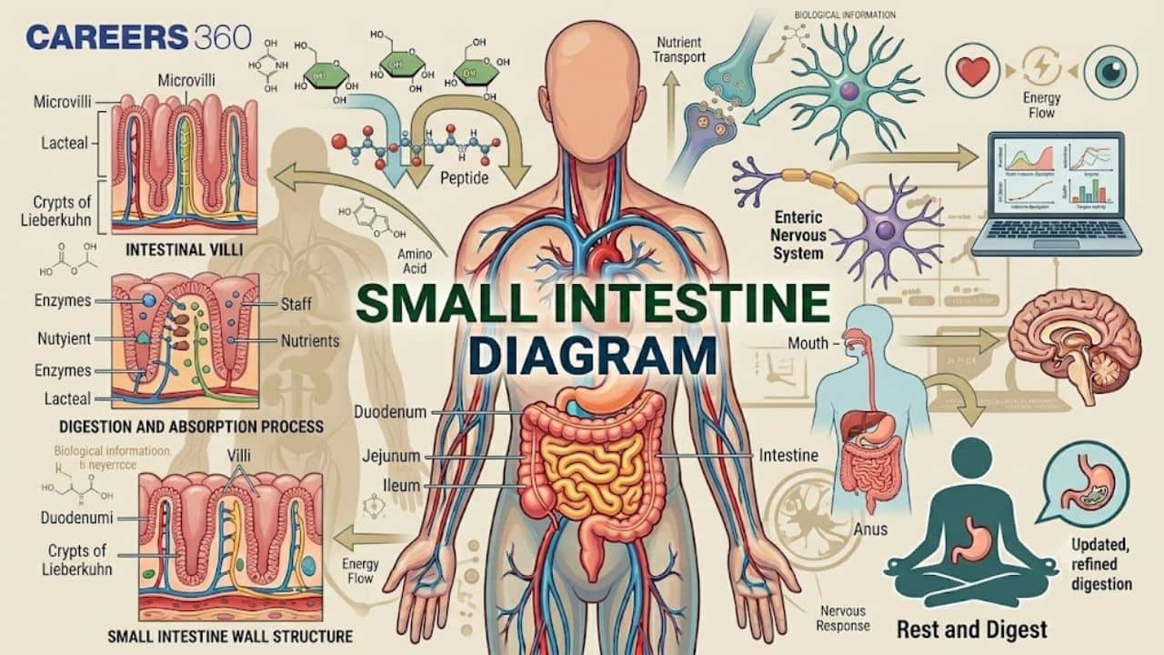
Small Intestine Diagram
A labelled diagram of small intestine shows its structure and functions in digestion and absorption. The parts of the small intestine diagram consist of the duodenum, jejunum, and ileum. These small intestine parts are essential for the digestion and absorption of food particles. The TS of the small intestine have specialised layers and villi for nutrient uptake. It helps students understand how the food is broken down and nutrients are absorbed. This guide explains the small intestine diagram, labels, layers, villi, intestinal glands, and NEET-oriented questions of human physiology.
This Story also Contains
- What is the Small Intestine?
- Diagram of Small Intestine Parts and Functions
- Small Intestine Structure and Their Role in Digestion
- Layers of the Small Intestine (Diagram Explanation)
- Diagram of Small Intestine Villi and Microvilli
- Intestinal Glands in the Small Intestine
- Difference Between Duodenum, Jejunum, and Ileum
- Small Intestine NEET MCQs (With Answers & Explanations)
- Recommended Video on Small Intestine
The TS of the small intestine diagram makes it easy to see the mucosa, submucosa, muscularis, and serosa clearly. Each layer has a role in nutrient absorption. The diagram of the small intestine also explains villi, intestinal glands, and blood supply in the human digestive system. The parts of the small intestine diagram help students remember the differences between the duodenum, jejunum, and ileum.The study of these small intestine diagrams strengthens human physiology preparation for competitive exams like NEET and AIIMS.
What is the Small Intestine?
The small Intestine is the main component of the digestive system, which absorbs nutrients. It is a long, coiled tube that starts from the stomach, ends in the large intestine, and extends to a length of about 20 feet. Its long length and the structure efficiently process and absorb the needed nutrition for the body's functions.
The small intestine has three parts: the duodenum, jejunum, and ileum. The small intestine breaks down food into nutrients with the help of digestive enzymes and absorbs those nutrients into the bloodstream. Such absorption ensures that the body receives the energy and necessary nutrients, hence being highly important.
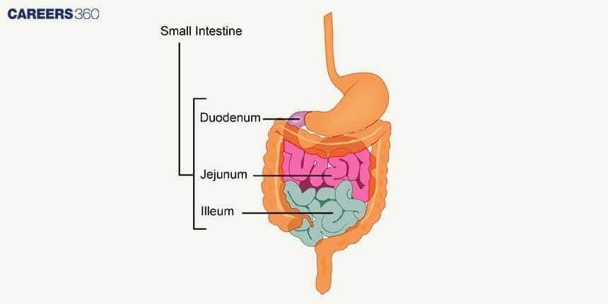
Diagram of Small Intestine Parts and Functions
The parts of the small intestine diagram highlight three main sections:
Duodenum - the first part, which receives bile and pancreatic juices.
Jejunum - the middle part, which absorbs most nutrients.
Ileum - the last part, which absorbs vitamin B12 and bile salts.
These small intestine parts work together to break down the food and absorb nutrients.
The various parts of the small intestine have different roles in digestion, breaking down food, and absorbing it into the bloodstream for utilisation.
Small Intestine Structure and Their Role in Digestion
The small intestine is mainly divided into three parts:
Duodenum
First part of the small intestine
Site for maximum chemical digestion.
Receives pancreatic juice and bile.
Jejunum
The middle portion.
Mainly responsible for the absorption of nutrients.
Rich blood supply.
Ileum
The last part.
Responsible for the absorption of vitamin B12 and bile salts.
Opens into the large intestine.
Layers of the Small Intestine (Diagram Explanation)
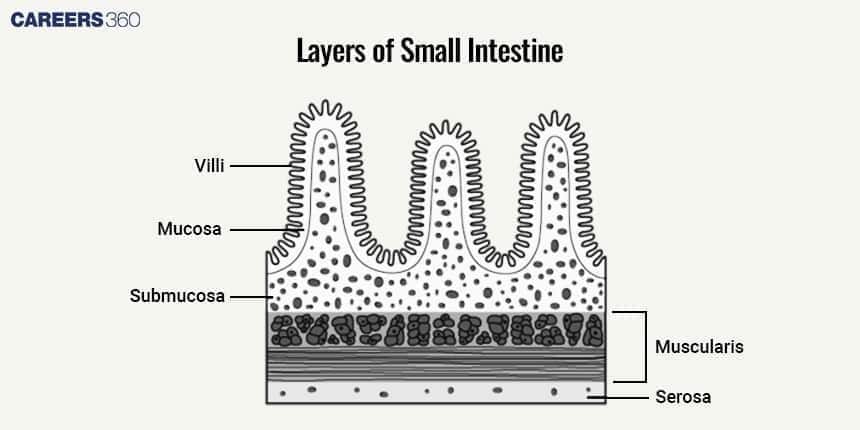
The layers of the small intestine are divided into:
Mucosa
The innermost layer.
Lined with villi and microvilli
It can increase the surface area for absorption.
Submucosa
A supportive layer containing blood vessels, nerves and glands.
Provides nourishment and support.
Muscularis externa
Circular and longitudinal muscles.
Responsible for the movements of peristalsis and segmentation.
Serosa
Outermost protective layer.
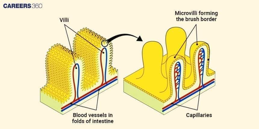
Diagram of Small Intestine Villi and Microvilli
The diagram of the intestinal villi represents:
Villi are finger-like projections.
Microvilli from the brush border.
Greatly increase surface area to promote nutrient absorption.
Blood vessels and lymphatics are found in these structures to transport the absorbed nutrients.
Intestinal Glands in the Small Intestine
Intestinal glands are located on the inner lining of the small intestine and are responsible for digestion. They secrete:
Digestive enzymes
Mucus
These glands are composed of different cells, including:
Goblet cells that produce mucus.
Enterocytes are involved in nutrient absorption.
Enteroendocrine cells that release hormones to regulate digestion.
Difference Between Duodenum, Jejunum, and Ileum
The small intestine is divided into three parts: the duodenum, the jejunum, and the ileum. Each section has a specific role in the digestion and absorption of nutrients. The difference between the small intestine's different parts is given below:
Aspect | Duodenum | Jejunum | Ileum |
Position | First part, just after the stomach | Middle part | Last part, before the large intestine |
Length | Shortest (~25 cm) | Medium (~2.5 m) | Longest (~3.5 m) |
Work | Digestion with bile and enzymes | Absorption of nutrients | Absorption of vitamin B12 and bile salts |
Feature | Brunner’s glands | Many folds and villi | Peyer’s patches (immune role) |
Small Intestine NEET MCQs (With Answers & Explanations)
Important questions asked in NEET from this topic are:
Compartment of the ruminant stomach
Digestion in Ruminants
Practice Questions for NEET
Q1. Identify the precise nature and origin of the enzyme Enterogastrone
An enzyme secreted by the gastric mucosa
A hormone secreted by the small intestinal mucosa
A hormone secreted by the duodenal mucosa
A secretory product of an endocrine gland associated with digestion
Correct answer: 3) A hormone secreted by the duodenal mucosa
Explanation:
Enterogastrone is a collective term for various hormones produced by the mucosa of the small intestine, primarily the duodenum. These hormones, such as secretin and cholecystokinin, are released in response to the presence of chyme in the small intestine and play a role in regulating digestive processes. The term "enterogastrone" specifically emphasizes its inhibitory effect on gastric motility and acid secretion, helping to slow down the emptying of the stomach and promote efficient digestion and absorption in the small intestine.
Hence, the correct answer is option 3) A hormone secreted by the duodenal mucosa.
Q2. Assertion: The serosa is a smooth membrane made up of a thin layer of cells that release serous fluid and a thin layer of connective tissue.
Reason: Serous fluid is a lubricating fluid that minimises friction caused by muscularis action
Assertion and reason are both true, and reason is an accurate account of assertion.
Both assertion and reason are accurate, but reason does not adequately explain assertion.
The assertion is correct, but the reasoning is incorrect.
Both the assertion and reason are incorrect.
Correct answer: 1) Assertion and reason are both true, and reason is an accurate account of assertion.
Explanation:
The serosa is the outermost layer of the small intestine and is composed of a thin layer of cells that secrete serous fluid and a layer of connective tissue. The serous fluid serves as a lubricant that reduces friction caused by the movement of the muscularis layer, which helps the small intestine mix and propel its contents along its length. Therefore, both the assertion and reason are true, and reason accurately explains the assertion.
Hence, the correct answer is option 1) Assertion and reason are both true, and reason is an accurate account of assertion.
Q3. Last section of large intestine is
Caecum
Colon
Rectum
Anus
Correct answer: 3) Rectum
Explanation:
The rectum is the final part of the large intestine, situated just before the anus. It plays a vital role in the elimination of waste from the body. The rectum stores formed faeces, which are the byproducts of digestion and absorption until they are ready to be expelled from the body during defecation.
Hence, the correct answer is option 3) Rectum.
Recommended Video on Small Intestine
Frequently Asked Questions (FAQs)
Common disorders include Crohn's disease, celiac disease, and irritable bowel syndrome.
In herbivores, it takes longer to help digest fibrous plant material. In carnivores, it is shorter due to their protein-rich diet.
The small intestine is divided into three parts: the duodenum, jejunum, and ileum.
The small intestines help in digestion by the activity of enzymes and bile breaking food into absorbable nutrients.
Villi are small, finger-like projections that increase the surface area for nutrient absorption in the small intestine.
