

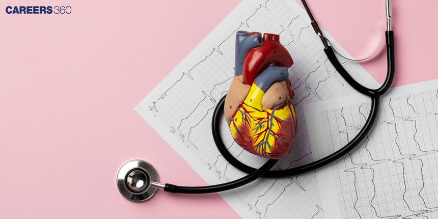
Regulation Of Cardiac Activity: Definition And Diagram
Regulation of cardiac activity involves intrinsic, neural, hormonal, and reflex mechanisms that maintain proper heart function. These processes ensure stable cardiac output, adequate tissue perfusion, and adaptation to physiological states like exercise, stress, and sleep. This guide covers mechanisms of regulation, cardiac cycle overview, reflex pathways, HRV, disorders, NEET MCQs, and FAQs.
This Story also Contains
- Overview of Cardiac Cycle
- Basic Anatomy and Physiology of the Heart
- Regulation of Cardiac Activity
- Neural Regulation (ANS Control of Heart)
- Hormonal Regulation
- Intrinsic Regulation – Frank-Starling Mechanism
- Reflex Mechanisms in Cardiac Regulation
- Regulation of Cardiac Output
- Heart Rate Variability (HRV)
- Cardiac Regulation in Different Physiological States
- Disorders Related to Cardiac Regulation
- Regulation of Cardiac Activity NEET MCQs (With Answers & Explanations)
- Recommended video on Regulation of Cardiac Activity
Overview of Cardiac Cycle
The cardiac cycle refers to the events occurring in a single heartbeat. It is the alternate contraction and relaxation of the chambers of the heart. It involves two major phases: systole, when the ventricles contract and blood is pushed out of the heart, and diastole when the heart muscle relaxes and allows the chambers to fill with blood. The effect of this cycle is an effective blood flow to the body, delivering needed oxygen and nutrients to tissues and taking away wastes.
Basic Anatomy and Physiology of the Heart
The anatomy of the heart is discussed below:
Location
The heart is a two-sided, four-chambered muscular pump positioned in the thoracic cavity, flanked by the lungs. Enclosed is a two-layered covering called the pericardium.
Chambers
The chambers are separated by walls of muscle. The two right and left atria are the upper chambers of the heart that receive the blood coming to the heart. The last two chambers of the heart are the ones that pump blood out of the heart, these are the right and left ventricles.

Regulation of Cardiac Activity
Cardiac activity is regulated by the sympathetic and parasympathetic of the autonomic nervous system. Hormones like adrenaline and thyroxine increase cardiac output during stress or activity. Baroreceptors and chemoreceptors modulate heart function in response to changes in blood pressure. Additionally, physical factors such as temperature and electrolyte balance also influence overall cardiac performance.

Neural Regulation (ANS Control of Heart)
Cardiac output variation and rhythmic control can also be provided by the autonomic nervous system ANS impulse generation. The ANS activity takes place in the brain's medulla oblongata. There exist two kinds of nerves in the system: the sympathetic nerves and the parasympathetic nerves, which are opposite to each other.
Sympathetic Control
The sympathetic nerves increase the strength of ventricular and atrial contraction when stimulated.
This will result in an increased cardiac output.
Heart rate also increased.
Parasympathetic Control
Compared to a sympathetic action, parasympathetic stimulation reduces the contraction of atria and ventricles.
Hence, giving less cardiac output and heart rate.
Hormonal Regulation
Apart from autonomic regulation, there are certain chemicals which can affect the regulation of cardiac activity. These chemicals include hormones like epinephrine, norepinephrine, and thyroxine.
Key Hormones
They can increase contraction and heart rate.
The other chemicals that impact the heart are ions.
Not only these factors but also the gender of an individual has an impact on the regulation of cardiac activity.
Intrinsic Regulation – Frank-Starling Mechanism
The Frank-Starling Law of the Heart describes an event where, when the volume of blood filling the heart increases, the stroke volume increases.
This intrinsic regulation implies that the more blood that comes back to the heart, the more the heart is going to pump out in a very nice balancing act for circulation.
Reflex Mechanisms in Cardiac Regulation
There are two main reflex mechanisms in cardiac regulation:
Baroreceptor Reflex
The baroreceptors, located within the carotid arteries and aorta, sense changes in blood pressure. The impulses that are impinged through it are transmitted to the brain, which responds by modifying the heart rate together with the diameter of the blood vessels, hence keeping the blood pressure stable.
Chemoreceptor Reflex
These chemoreceptors are found in the carotid bodies and aorta. They establish reflexes responding to alterations in blood gases, primarily carbon dioxide and oxygen. Those reflexes, via these receptors, play a huge role in the regulation of respiration and cardiovascular activities.
Regulation of Cardiac Output
Cardiac output refers to the volume of blood pumped by the heart in one minute. It is determined by heart rate and stroke volume.
Factors Influencing CO
The factors that influence cardiac output are venous return, peripheral resistance, and the strength of contraction of the ventricles.
The cardiac output must be maintained at a level sufficient to provide adequate blood supply to all tissues.
Heart Rate Variability (HRV)
Heart rate variability is the measure of time variation between each heartbeat and acts as an index for sympathetic-parasympathetic balance. HRV is an important index for cardiovascular health and autonomic nervous system function.
The methods physically offered for the measurement and techniques of HRV include time-domain, frequency-domain, and nonlinear analyses.
Cardiac Regulation in Different Physiological States
Cardiac regulation adapts to different physiological conditions:
During exercise, there is an increase in heart rate and stroke volume as an attempt to meet the oxygen demand of muscles, which is higher.
In response to stress, the sympathetic nervous system is activated, and heart rate and cardiac output increase.
The parasympathetic dominance is the sleep stage, during which the heart rate is lower.
Ageing may affect cardiac regulation; there is a general decline in cardiac output and a predisposition to arrhythmias.
Disorders Related to Cardiac Regulation
Several disorders are associated with impaired cardiac regulation:
Hypertension: This may be a constant straining of the heart that could cause intermittent injuries in blood vessels over time.
Arrhythmias: These are irregularities in the heartbeat, which prevent the smooth flow of blood and can lead to severe consequences.
Heart Failure: The inability of the heart to pump blood effectively could be due to a weakening of the cardiac muscle or some defect in the structure.
Chronic Diseases: Conditions such as diabetes mellitus and hyperthyroidism are also known to affect the heart and its regulations.
Regulation of Cardiac Activity NEET MCQs (With Answers & Explanations)
Types of questions asked from this topic are:
Regulation of cardiac activity
Disorders related to cardiac activity
Practice Questions for NEET
Q1. Which among the following is correct during each cardiac cycle?
The volume of blood pumped out by the Rt and Lt ventricles is the same
The volume of blood pumped out by the Rt, and Lt ventricles is different.
The volume of blood received by each atrium is different.
The volume of blood received by the aorta and pulmonary artery is different.
Correct answer: 1) The volume of blood pumped out by the Rt and Lt ventricles is the same
Explanation:
After oxygenation, the volume of the blood entering the lung is the same as the volume of blood leaving it. In the case of varying volumes, the heart is susceptible to heart failure due to different pressures. If the volume of blood entering the lungs exceeds the capacity for oxygenation or drainage, it can cause fluid buildup, leading to pulmonary congestion. This imbalance in pressure can strain the heart, ultimately contributing to heart failure if left unaddressed.
Hence, the correct answer is option 1) The volume of blood pumped out by the Rt and Lt ventricles is the same.
Q2. Using which information can the amount of blood ejected from the left ventricle over a minute be calculated?
Heart rate and the fluid volume drunk on the day
Stroke volume and Circulating blood volume
Circulating blood volume and the fluid volume drunk on the day
Stroke volume and heart rate
Correct answer: 4) Stroke volume and heart rate
Explanation:
Stroke volume is the amount of blood ejected from the ventricle with each cardiac cycle. It can be readily calculated by subtracting the end-systolic volume from the end-diastolic volume. Multiplying the stroke volume by the heart rate yields the cardiac output, typically reported in liters per minute. A normal resting heart rate for adults ranges from 60 to 100 beats per minute.
Generally, a lower heart rate at rest implies more efficient heart function and better cardiovascular fitness. For example, a well-trained athlete might have a normal resting heart rate closer to 40 beats per minute.
Hence, the correct option is 4) Stroke volume and heart rate.
Q3. Which of the following definitions accurately describes cardiac output?
The amount of blood accumulated in the atrium before contraction.
The quantity of blood gathered in the ventricles during relaxation.
The volume of blood propelled from the atrium to the ventricles during contraction.
The quantity of blood expelled from the ventricles into the aorta and pulmonary artery.
Correct answer: 4) The quantity of blood expelled from the ventricles into the aorta and pulmonary artery.
Explanation:
Cardiac output refers to the amount of blood pumped out of the ventricles of the heart per unit of time, typically measured in liters per minute. It represents the volume of blood ejected from the ventricles and delivered into the systemic circulation (aorta) and pulmonary circulation (pulmonary artery). Therefore, option 4 accurately describes cardiac output.
Hence, the correct answer is option 4) The quantity of blood expelled from the ventricles into the aorta and pulmonary artery.
Also Read:
Recommended video on Regulation of Cardiac Activity
Frequently Asked Questions (FAQs)
A raised blood pressure, first detected by baroreceptors, provides negative feedback that leads to reflexes that slow the heart and dilate blood vessels, thereby returning blood pressure to normal.
HRV measures the variation in time between heartbeats and reflects autonomic nervous system activity, a major determinant of overall cardiovascular health.
The ANS controls heart rate and the force of contraction through its two divisions: the sympathetic and parasympathetic divisions.
Adrenaline and thyroid hormones are some of these hormones, which increase heart rate and cardiac output by acting directly on cardiac cells to change their activity.
The Frank-Starling Law states that stroke volume increases when there is more blood volume within the heart.
