

Thorax: Definition, Function, Diagram and Examples
The thorax is the central part of the body located between the neck and abdomen. It forms the thoracic cavity. It houses vital organs such as the lungs, heart, oesophagus, and major blood vessels. The rib cage, sternum, and thoracic vertebrae protect these organs. The thorax plays an important role in respiration, circulation, and structural support.
This Story also Contains
- What is the Thorax?
- Anatomy Of The Thorax
- Organs Present in the Thorax
- Functions of the Thorax
- Thorax Diagram
- Clinical Importance of Thorax
- Thorax NEET MCQs (With Answers & Explanations)
- Recommended Video on "Thorax"
The anatomy of the thorax explains how bones, muscles, and organs work together to maintain life processes. The diaphragm and intercostal muscles enable breathing, while the heart and vessels ensure blood circulation. The thorax cavity also provides a structural framework for the upper body and supports muscle attachment. Studying the thorax definition, functions, and examples is essential for NEET, nursing, and medical entrance exams.
What is the Thorax?
The thorax is the body region located between the neck and abdomen. It accommodates the heart and lungs, though they are shielded by the rib cage of the human body. In humans and many other organisms, the thorax is of great significance because it introduces a cavity that facilitates the movements of respiratory organs in respiration as well as the movements of circulatory organs in circulation. To summarise, it is crucial for the stability, as well as the functionality of the respiratory and cardiovascular systems. Thus, it occupies a rather crucial place in both human and animal physiology.
Anatomy Of The Thorax
The thorax is the region between the neck and abdomen. It forms the thoracic cavity, which protects and supports vital organs. The anatomy includes bones, muscles, and internal structures that work together for respiration and circulation. The structure of the thorax has the following components:
Thoracic Skeleton
The thoracic cavity is formed by the thoracic part of the vertebral column, sternum, and ribs. It encases the heart and lungs. The sternum is smooth at the bottom and linked to the ribs through cartilage. The ribs are connected to the thoracic vertebrae at the back.
Thoracic Muscles
Some of them are intercostal muscles that help in respiration as well as contraction of the chest, and the diaphragm, which has a significant role in increasing the volume of the thoracic cavity. The muscles are the scalene and the sternocleidomastoid, which assist in deep breathing.
Thoracic Cavity
The thoracic cavity is a part of the body that is enclosed by the rib cage, sternum and thoracic vertebrae. It is subdivided into two pleural cavities containing the lungs and the mediastinum, which contains the heart, oesophagus and trachea.
Organs Present in the Thorax
The thorax houses vital organs essential for respiration, circulation, and digestion. These include the lungs, heart, oesophagus, and major blood vessels. Together, they maintain life processes and protect body fluid and circulation stability.
Lungs
The lungs are the primary organs of respiration. They enable gas exchange by absorbing oxygen and releasing carbon dioxide.
Heart
The heart pumps blood throughout the body. It maintains circulation and supplies oxygenated blood to tissues.
Oesophagus
The oesophagus is a muscular tube that connects the throat to the stomach. It transports food and liquids through peristalsis.
Major Blood Vessels
The thorax contains the aorta, vena cava, and pulmonary vessels. These regulate blood flow between the heart, lungs, and body.
Functions of the Thorax
The thorax plays a vital role in respiration, circulation, and the protection of organs. It provides a strong structural framework and supports survival by multiple systems. The functions of the thorax are:
Protection of Vital Organs
The thorax safeguards structures like the heart, lungs and the majority of blood vessels against physical harm. The rib bones and sternum create a strong armour that effectively protects the injured. The thoracic vertebra also supports the spinal column as well as the nerve trunks.
Role in Respiration
For breathing, one has to use the thorax to expand while taking in air and contract while expelling the air. The diaphragm becomes flattened on contraction, lifts the lower ribs and enlarges the thoracic cavity, thus creating a vacuum that pulls the air into the lungs. Since breathing entails the movement of air in and out, the intercostal muscles also help in lifting and lowering the ribs.
Circulatory Support
The thorax was also concerned with the circulation of blood since it contains the heart and large vessels, including the aorta and pulmonary arteries. Thoracic movement during respiration also facilitates the return of the blood in the veins to the heart and promotes proper blood circulation in the thoracic cavity.
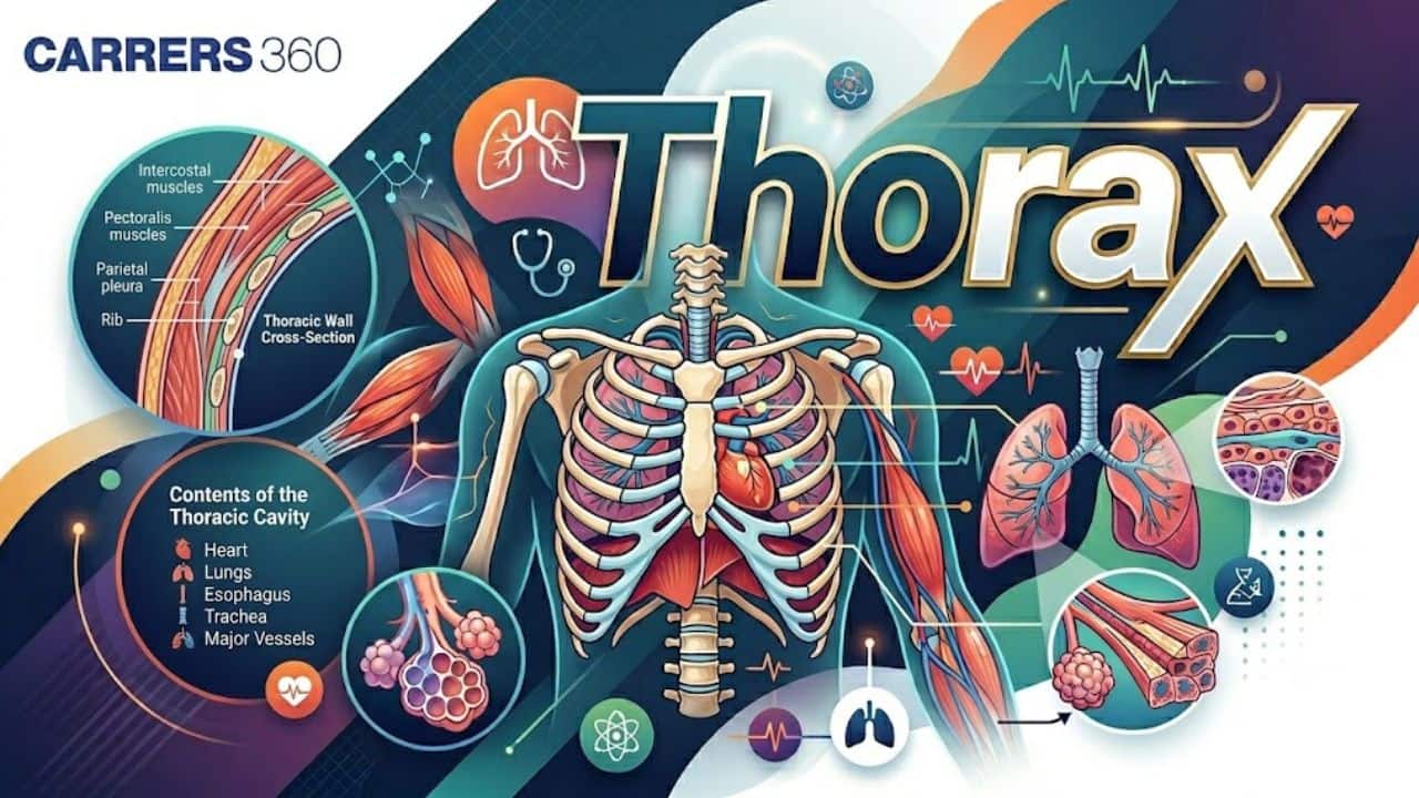
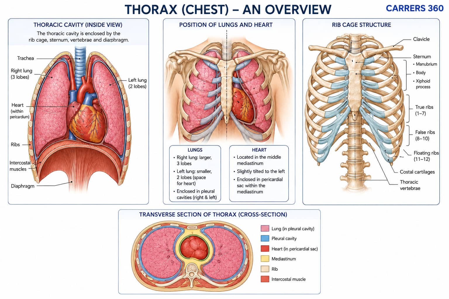
Thorax Diagram
The thorax is a protective bony cage formed by the ribs, sternum, and thoracic vertebrae. It houses the lungs and heart, protects these vital organs, and assists in breathing through the movement of the ribs and rib cage with the diaphragm.
Clinical Importance of Thorax
The thorax is clinically important because it houses vital organs like the lungs, heart, oesophagus, and major blood vessels. Any injury or disease in this region can affect respiration, circulation, and digestion. The thorax can have these issues:
Thoracic Injuries
Thoracic injuries, which include the ribs or any blunt force, pose a risk to the organs within the thoracic cavity, which include the lungs and heart. Broken ribs can be painful and hinder the patient’s ability to breathe, while severe injuries may cause internal bleeding or affect their breathing.
Chest Pain Causes
Most people experience chest pain in the thoracic region from numerous causes like costochondritis, which is a non-serious condition, but also from serious conditions like myocardial infarction or pulmonary embolism. They can be described as sharp, dull or pressurising pain and usually cannot be distinguished until further investigations are done to establish the cause.
Atelectasis
Atelectasis means the lung or a certain lobe does not inflate as it should, possibly resulting from obstruction or pressure injury. It can occur due to pneumonia, tumour or after an operation, and it causes less oxygenation and breathing problems.
Pneumothorax
Pneumothorax, on the other hand, is caused by the entrapment of air in the pleural cavity, hence making the lung collapse. It may be caused by trauma, medical treatment, or development on its own. Thus causing sharp chest pain and difficulty breathing with difficulty. Management might entail the aspiration of the air and allow the lung to reinflate.
Thorax NEET MCQs (With Answers & Explanations)
Important topics for NEET are:
Functions and anatomy of thorax
Clinical aspects of Thorax
Practice Questions for NEET
Q1. Which of the following statements about the pleural cavity is incorrect?
It is a potential space between the visceral and parietal pleura.
It is filled with pleural fluid.
It helps reduce friction during breathing.
It is located within the mediastinum.
Correct answer: 4) It is located within the mediastinum
Explanation:
The mediastinum does not contain the pleural cavity. The heart, major vessels, esophagus, and trachea are all located in the mediastinum, which is the thoracic cavity's core compartment. On the other hand, the visceral and parietal pleura, two layers of serous membrane that encircle the lungs, may be separated by the pleural cavity. Pleural fluid, which fills the pleural cavity, serves to lessen friction while breathing and enables the lungs to expand and contract without resistance.
Hence, the correct answer is option 4) It is located within the mediastinum.
Q2. Oblique fissure is present in
Left lung
Right lung
Both
None
Correct answer: 3) Both
Explanation:
The right lung consists of three lobes—superior, middle, and inferior—separated by two fissures: the horizontal fissure (between the superior and middle lobes) and the oblique fissure (between the middle and inferior lobes). In contrast, the left lung has only two lobes—superior and inferior—divided by a single oblique fissure. The left lung is slightly smaller than the right lung due to the space occupied by the heart, and it lacks the middle lobe that is present in the right lung.
Hence, the correct answer is option 3) Both.
Q3. Where are lungs situated in the human body ?
Abdominal cavity
Thoracic cavity
Coelomic cavity
Pleural cavity
Correct answer: 2) Thoracic cavity
Explanation:
The lungs are situated in an air-tight chamber called the thoracic chamber. The thoracic chamber is formed dorsally by the vertebral column, ventrally by the sternum, laterally by the ribs, and on the lower side by the dome-shaped diaphragm.
Hence, the correct answer is option 2) Thoracic cavity.
Recommended Video on "Thorax"
Frequently Asked Questions (FAQs)
The thorax is the organ of the heart, lungs, gullet, trachea and major blood vessels such as the aorta and pulmonary arteries.
The rib cage and the sternum that begins at the base of the neck cup the highly vulnerable heart and lungs and shield them from different physical impacts.
The limbs that make up the thorax include; it is mainly comprised of four limbs that are vital for protecting vital body organs, and circulation since they hold the heart and major vessels while the lungs for respiration.
The mammal will have a rib cage as well as a diaphragm comparable to humans. However, birds and reptiles have different ribs and respiratory muscles for their breathing styles.
Other ordinary illnesses and factors are rib break, pneumonia, atelectasis, pneumothorax as well as thoracic pain resulting from costochondritis or myocardial infarction.
