

Simple Microscope - Definition, Diagram, FAQs
A simple microscope is a basic optical instrument used to view small objects as enlarged images using a single convex lens. It works on the principle of magnification by forming a virtual, erect, and magnified image when the object is placed within the focal length of the lens. Also known as a magnifying glass, the simple microscope was first developed by Antonie van Leeuwenhoek and laid the foundation for modern microscopy. Understanding the simple microscope definition, parts, working principle, magnification formula, and uses is important for students studying Class 12 Physics. This topic is frequently asked in NCERT exams and competitive entrance tests. Simple microscopes are widely used in laboratories, biology, medicine, and everyday applications.
What is a Simple Microscope?
Simple Microscope Definition: A Simple Microscope meaning is used to see a magnified image of an object. Antonie Van Leeuwenhoek, a Dutchman, invented the first simple microscope, consisting of a single powerful magnetic lens that rotates to detect tiny freshwater insects. It is composed mainly of light microscopes. The main property of the target lens used in microscopes is to produce a virtual, upright, and magnified image when the object is placed within a fixed height. A convex lens used in a microscope is to create a simple microscope. Convelenses are widely used as a reading glass or magnifying glass. Now, to obtain high magnification, a combination of two or more convex lenses is used to create an integrated microscope.
What Are the Parts of a Simple Microscope?
A Simple Microscope contains various optical components and other supporting (or mechanical) components as discussed in this section, the layout of a simple basic microscope with its various labeled numbers. The Eyepiece is connected to the lens via a tube. The eyeball is the lens where the image of the object can be seen. Tube length varies by rotating the button so that a clear image is obtained by changing the focus. The objective lens enhances magnification.
A simple Schematic of the Magnifying power of a compound microscope showing its different parts.
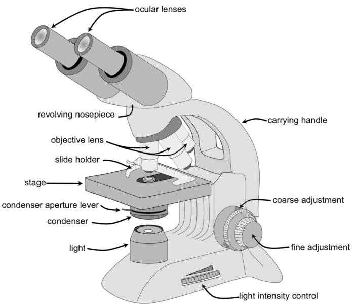
The sample platform, made of metal foil, consists of metal clips holding a sample, which is placed on a glass slide, under observation. The mirror focuses on the light of the sample. All components are grounded.
The base is a component of equipment that provides support for the capture of other parts of the microscope. The arm of the microscope is connected to a physical object.
The following are the components of a Simple Microscope and their functions:
Eyeball: A lens used to study samples and placed on top. It has an increase of 10X to 15X.
Baseline: This provides microscope support.
Tube: This is used to connect an eyepiece to the target lens.
Target lenses: These are available in 10X, 40X, and 100X magnification lenses and have color coding. The lower electric lenses are shorter lenses and the upper electric lenses are much longer.
A flexible nose piece: This is also known as a turret. It is used to capture other purpose lenses and can be changed while viewing samples.
Diaphragm: Used to control the amount of light passing through the stage.
Stage: It is a platform used to place slides with samples.
Stage Clip: This is used to hold slides in the right place.
Adjust location button: Used to focus on scanning.
Good repair box: Used to focus on oil.
Arm: Used to support the tube and connect to the base of the microscope.
Power switch: A primary power switch used to turn the microscope on or off.
Condenser: Used to focus the light on the sample and uses 400X electric lenses.
Magnification of a Simple Microscope
It is defined as the ratio of the angle subtended by the image at the eye to the angle subtended by the object when placed at the least distance of distinct vision.
Case 1: Image at Infinity
$M=\frac{D}{f}$
Case 2: Image at Least Distance of Distinct Vision (D)
$
M=1+\frac{D}{f}
$
Where:
- $M=$ magnification
- $D=$ least distance of distinct vision $(\approx 25 \mathrm{~cm})$
- $f=$ focal length of the convex lens
Working of Simple Microscope
A simple microscope consists of a single convex (biconvex) lens and is used to obtain a magnified view of small objects. The object to be observed is placed within the focal length of the lens. When light from a source falls on the object, it gets transmitted or reflected and enters the convex lens.
The lens bends (refracts) the light rays in such a way that they appear to come from a larger distance, forming a virtual, erect, and magnified image on the same side of the lens. For maximum magnification, the object is kept very close to the lens, and the final image is formed either at the least distance of distinct vision or at infinity.
Proper illumination and contrast can be achieved by adjusting the intensity of light, often using a diaphragm. In biological applications, staining is used to enhance visibility of fine details.
Simple Microscope Experiment
Aim
To construct a simple microscope using a water droplet and observe magnification.
Apparatus Required
- A glass of water
- Fuse wire
- Object to view (e.g., newspaper with fine print)
Procedure
- Make a small loop (about 2 mm wide) using the fuse wire.
- Dip the loop into water so that a small droplet forms in it.
- Hold the loop close to your eye.
- Place the object near the water droplet and adjust the distance.
- Observe the image formed through the droplet.
Observation
A magnified and clear image of the object is seen when the droplet is held at an उचित distance.
Conclusion
The water droplet acts like a convex lens, refracting and converging light rays to produce a magnified image. This principle is similar to early microscopes, where small glass or water globules were used for magnification. Modern microscopes use multiple precisely shaped lenses to observe very fine details.
Difference Between Simple and Compound Microscope
| Basis | Simple Microscope | Compound Microscope |
| Definition | Uses a single convex lens for magnification |
Uses two or more lenses (objective + eyepiece) |
| Magnification | Low (generally up to 10–20×) | High (can be 100× to 1000× or more) |
| Construction | Simple and compact | Complex with multiple optical parts |
| Image Formation | Single virtual, erect image |
Final image is highly magnified and virtual (initial image is real and inverted) |
| Usage | Used for reading, magnifying small objects |
Used in laboratories to observe cells, microorganisms |
| Resolution | Low | High |
| Cost | Low | Expensive |
Use of a Simple Microscope
- Used in pedology (study of soil particles)
- It is used by a dermatologist to diagnose various skin diseases.
- It is used in microbiology to study algae samples, fungi, etc.
- It is used by carpenters to obtain an enlarged view of fine jewellery.
Also check-
- NCERT Exemplar Class 11th Physics Solutions
- NCERT Exemplar Class 12th Physics Solutions
- NCERT Exemplar Solutions for All Subjects
NCERT Physics Notes:
Frequently Asked Questions (FAQs)
A Simple Microscope contains a single lens that is traditionally called a loupe. A well-known example today is the reading or magnifying glass. Modern high-resolution lenses are usually made of two-dimensional glass materials that produce a colour-coded image.
A convex lens is used to create a simple microscope. A convex lens is widely used and widely used as a reading glass or magnifying glass.
Examples of simple microscopes include reading glasses, decorative ornaments, and pocket magnifiers. ... There are two types of integrated microscopes that use light to detect visible (or flexible) objects and invisible objects. An opaque object requires reflective light from an object through the optic tube lens.
Converting the lens is called a convex lens that converts the rays of the light of the event to the same as the main axis at the same time; this is why flexible lenses are used in objects such as microscopes to focus on minute particles.
In the microscope, we often use a convex lens because the convex lens even magnifies images. Microscopes produce highly enlarged images of very small objects for this purpose the convex app is very useful. ... However, between the three lenses, the lens located at the end of the microscope produces a transformed and enlarged image.
