


Structures in Bacteria: Definition, Diagram, Classification, Microbes, Cells
Bacteria are unicellular and prokaryotic microorganisms. They lack a true nucleus and membrane-bound organelles, but have a simple cellular organisation that helps them to survive in diverse environments. Bacteria are found in soil, water, air, extreme habitats, and even inside the bodies of plants and animals.
This Story also Contains
- What Is Bacteria?
- Classification Of Bacteria
- Differences Between Gram-positive and Gram-negative Bacteria
- Basic Bacterial Cell Structure
- Cytoplasmic Structures
- External Structures
- Specialised Structures
- Intracellular Inclusions
- Recommended video on Structures in Bacteria
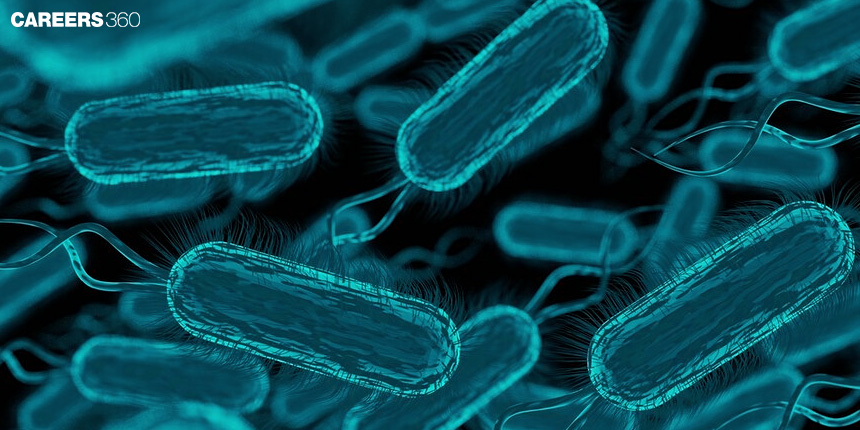
Bacteria play a very important role in nutrient cycling, nitrogen fixation, decomposition, food production, etc. While many bacteria are beneficial, some are pathogenic and cause diseases. Understanding the basic bacterial structure is important for Class 11 Biology and is asked directly in competitive exams such as NEET.
What Is Bacteria?
Bacteria are single-celled microorganisms that belong to the domain prokaryote since they do not have a true nuclear membrane. Bacteria are present just about everywhere, including in hot hydrothermal vents, and the human gastrointestinal tract. Hence, bacteria are important in the different ecosystems since they are known to contribute to nutrient cycling, decay of organic material and easing the balance of ecosystems. They are essential in human health as commensal organisms that help in digestion and as agents involved in biotechnological uses like antibiotic synthesis and fermentation. But there is another group of bacteria that can be pathogenic, and become carriers of diseases that threaten society.
Classification Of Bacteria
Bacterial classification is discussed below
Based On shape
Based on shape: From the above classification, the bacterial cells are classified by their shapes as:
Cocci – spherical
Bacilli – rod-shaped
Vibrios – comma-shaped
Spirilla – spiral-shaped
Based on Gram staining
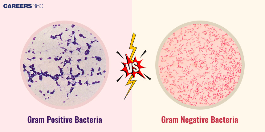
Based on Gram staining: Be converted into two groups; the first category is called Gram-positive which is the one that retains the crystal violet dye and the second category is called Gram-negative which does not retain the dye.
Other classifications
According to the bacterial oxygen requirements, the bacteria can be again divided into aerobic, which require oxygen for their survival, anaerobic, which do not survive in the presence of oxygen, and facultative anaerobes, which can survive in both conditions.
Differences Between Gram-positive and Gram-negative Bacteria
Here are a few differences between gram-positive and gram-negative bacteria:
Feature | Gram-positive Bacteria | Gram-negative Bacteria |
Gram stain result | Retain the crystal violet stain and appear purple | Do not retain crystal violet; appear pink/red after counterstaining |
Peptidoglycan layer | Thick | Thin |
Outer membrane | Absent | Present |
Teichoic acids | Present | Absent |
Examples | Staphylococcus, Streptococcus, Bacillus | Escherichia coli, Salmonella, Vibrio cholerae |
Basic Bacterial Cell Structure
The cell structure is discussed below:
Cell Wall
It has a thick layer of peptidoglycan in the cell wall and when stained with gram, the purple dye, crystal violet, is retained hence referred to as gram positive. On the other hand, the cell wall of the gram-negative bacteria has a relatively thinner peptidoglycan layer and is located between the inner and the outer membrane that contains lipopolysaccharide in the outer membrane part which makes them differ when stained with gram stain.
Functions Of The Cell Wall
Their structure has the support of the cell wall and protection from pressure changes in osmotic concentration. It also acts as a protective shield for the bacterial cell against unwanted materials and forms part of the structure and firmness of the bacterial cell.
Diagram of Gram-positive vs. Gram-negative bacteria
Plasma Membrane
The outer layer of the bacterial cell includes a plasma membrane consisting of a phospholipid bilayer with proteins integrated. The plasma membrane is a selectively permeable phospholipid bilayer that regulates the movement of substances into and out of the cell while maintaining the internal cellular environment.
Role In Nutrient Transport And Cell Signalling
It allows the transport of nutrients into the cell and the removal of waste products. Some molecular transport across Bacterial membranes occurs through membrane proteins, and about signalling, there are receptors on the bacterial membrane that give the bacteria the ability to note changes in environments leading to phenomena such as Chemotaxis and Quorum sensing.
Cytoplasmic Structures
The cytoplasmic structures are listed below
Cytoplasm
The cytoplasm is a gel-like matrix containing ribosomes, enzymes, metabolites, inclusion bodies, and the nucleoid region. Since bacteria are prokaryotes, they lack membrane-bound organelles.
Role In Metabolic Activities
Biomolecules are assigned to catalyse biochemical reactions including glycolysis, the citric acid cycle and synthesising amino acids and nucleotides in bacteria to produce energy and regulate their cellular processes.
Nucleoid
The nucleoid in bacteria is an area found in the cytoplasm of the bacteria that has a single circular chromosome. Hence while human cells contain genetic DNA this DNA is not contained within a membrane-enclosed nucleus but is more structured and condensed by proteins. For these and other purposes, the nucleoid also contains proteins that are involved in the processes of DNA replication transcription and repair.
Differences from the eukaryotic nucleus
While the eukaryotic nucleus is surrounded by a membrane and has multiple linear chromosomes the nucleoid of bacteria is not membrane surrounded. It is negative for histones and introns and its hereditary materials are characteristically shorter and denser. The nucleoid is also involved in bacterial cell division where through the breakdown of the cell the replicated DNA is divided between the daughter cells.
Ribosomes
Structure and function in protein synthesis
Ribosomes are cell organelles manufactured from RNA and protein. The ribosome is of two types – prokaryotic and eukaryotic – The prokaryotic ribosome is smaller in size than the eukaryotic ribosome. These include a big and a small portion of the ribosome which combine during translation. Ribosomes are very important structures that synthesize proteins through the process of translation, in which the mRNA strand codes for amino acid sequences that in turn fold into functional proteins. Bacterial ribosomes are 70S ribosomes composed of 50S and 30S subunits. They are the target of several antibiotics such as tetracycline and streptomycin.
External Structures
The external structures are listed below
Flagella
Flagella are thin structures that protrude from the bacterial cell surface and usually resemble a whip. The bacterial flagellum rotates using the proton motive force, enabling bacterial movement (motility).
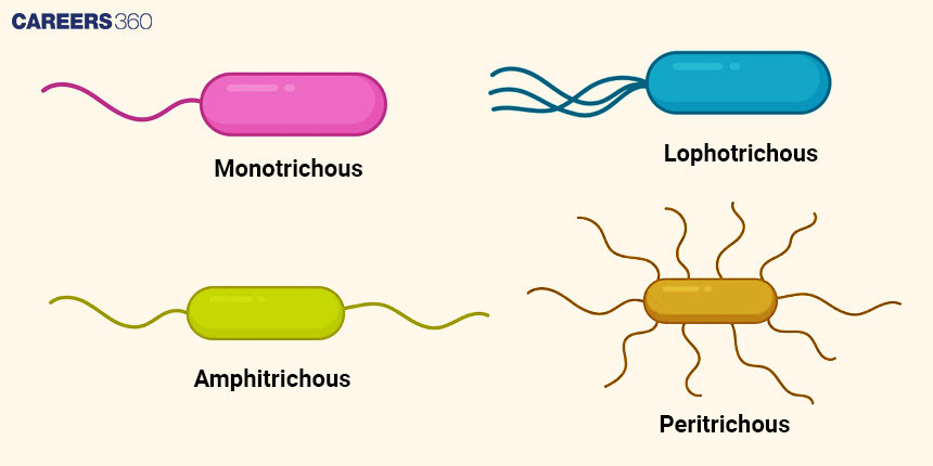
Types Of Flagellar Arrangements
Monotrichous: This showed only one flagellum located at the end of the cell.
Lophotrichous: Conclusions All flagella at one pole of the cell or multiple flagella at one end of the cell.
Amphitrichous: Flagella present in a single number and localized at both ends of the cell.
Peritrichous: Flagella present all over the surface of the cell
Diagram showing different flagellar arrangements
Pili and Fimbriae
Pili and fimbriae are short, hair-like protein appendages present on the surface of many bacteria. They are composed of the protein pilin.
Role In Adhesion And Conjugation
Fimbriae and pili allow bacteria to attach themselves to a surface host cell, or another bacterium. These adhesions play a very significant role during colonisation, biofilm formation and the infection processes.
Pili are also used in conjugation in which DNA (plasmids) is transferred from one bacterial cell to another. Conjugative pili are the structures or tubes which help in the process wherein the donor cell comes very close to the recipient cell and codes that can render resistance to antibiotics or any other positive quality are transferred.
Capsule And Slime Layer
Both capsules and slime layers are external layers of polysaccharides (and in some cases proteins) that surround the bacterial cell wall.
Differences Between Capsule And Slime Layer
Composition: Capsules are layers that are well compacted and close together, while slime layers are individual layers that are not very much in contact with each other.
Function: Capsules shield the bacteria from phagocytosis and drying, while slime layers are useful in holding the bacteria to surfaces and obtaining nutrients.
Importance In Bacterial Virulence And Protection
Capsules increase bacterial virulence by protecting bacteria from phagocytosis, preventing desiccation, and helping them adhere to host tissues. The slime layers assist bacteria in surviving in unfriendly surroundings and combat the antimicrobial opponents.
Specialised Structures
The specialised structures are listed below
Endospores
Endospores are obtained when bacteria go through sporulation since it is a method of surviving unfavourable conditions. They include the DNA body which is heavily compressed and packaged with the spore coat and cortex surrounding it.
Function In Bacterial Survival Under Harsh Conditions
Endospores allow bacteria to be in extreme conditions such as high temperature regimes or lack of humidity and chemical treatments and withstand them because the genetic material and the rest of the cell structures are protected.
Plasmids
Plasmids are small circular, self-replicating pieces of DNA that occur in addition to the bacterial chromosome. It can self-multiply within the bacterial cell and is common in conjunction with chromosomal DNA.
Role In Antibiotic Resistance And Genetic Variation
Thus, plasmids are essential in bacterial adaptation and existence at large. These organisms often hold genes that code for traits that enable bacteria to become resistant to antibiotics and their impact. Such genes can be of metabolic activities, toxins, or other factors making bacteria competitive and suitable for survival in given conditions carried by the plasmids.
Intracellular Inclusions
The intracellular structures are discussed below:
Inclusion Bodies
Inclusion bodies are diverse structures within bacterial cells that serve various functions
Glycogen granules: Used to store glucose as an emergency energy store during the availability of the nutrients to be used in emergencies when the nutrients are inadequate.
Gas vesicles: Supply floatation to control the position of the bacterium in water to ensure adequate aeration and irradiation.
Importance In Nutrient Storage
In other aspects, the inclusion bodies serve as nutrient granules where glycogen is stored thus allowing a period of starvation or stress in the bacteria. Gas vesicles help in improving the correct positioning in the environment for efficiency in the uptake of energy and use.
Recommended video on Structures in Bacteria
Frequently Asked Questions (FAQs)
In gram-positive bacteria, there is a thick layer of peptidoglycan and the absence of an outer membrane whereas in gram-negative there is a thin layer of peptidoglycan with the inner and outer membranes that contain lipopolysaccharide coating.
Flagella are appendages in bacterial cells, or filaments made of flagellin protein, transport in bacterial cells is driven by proton motive force with rotary movement. Eukaryotic flagella are slender threadlike structures that are composed of microtubules, organisational subunits of the cell supported by ATP and move in a whiplike manner for cell moving.
Endospores can be described as an exogenous resting structure produced in specific circumstances by certain bacteria. They conserve the bacterial DNA and the other important parts from degradation, thus, letting the bacterium withstand various conditions such as high temperature, dryness, and chemical solutions.
Both pili and fimbriae are protuberances that allow bacteria to attach to host tissues hence colonising and infecting them. They also regulate biofilm production which helps bacteria to survive in the host environment and resist.
They replicate independently and can contain other genes associated with characteristics that include antibiotic resistance, pathogenicity, or other metabolic aspects. They can self-propagate and be transferred from bacterium to bacterium; play a part in genetic variation and evolution.